The Amateur Mycologist - An Exploration Of Bizarre Sub-Millimeter Fungi And Insects Living In A Micro Ecosystem
These posts are not for foraging. They are intended for entertainment and intellectual satisfaction only. These posts are not a field guide nor comprehensive in any way - their accuracy is not assured in any way. Do not eat wild mushrooms unless you are a professional, have substantial professional assistance or have a wealth of personal experience with a specific species. Do not make any foraging decisions based on these posts. To do so could be dangerous or life threatening.
This post makes no reference to edibility or toxicity
These strange organisms are absolutely tiny, physically bizarre, and very enigmatic
Getting the photos of them that you see in this post took hours of prodding, twisting, lighting and post-processing. I completed the process about a week ago, and for the first time ever posted the information on my external blog before steemit in order to more easily be able to share the information with several specialized online communities.
I've posted these photos and the charactistics of these little sporangia to the Slime Mold Collective, the NYMS, and two subreddits specializing in fungi and slime molds respectively. I've read through several field guides in search of an answer, bought a copy of "The Kingdom Of Fungi" in the hopes of clarity, and then scoured more online sources than I'm going to cite to below.
In the end, there are certain informed, broad conclusions I can form about these organisms, but not many specific ones. This is likely to be a problem for me going forward at this scale. The fact seems to be that the more myopically you zoom in and search for tiny myco/myxo species, the less information is readily available about what you find.
There may be a mid to long term answer to this dilemma. The NYMS is going to be joining the North American Mycoflora Project, which will be encouraging amateurs to collect, document and send specimens for DNA testing. I will be attending an NYMS meeting on the topic in late January and, in time, hope to learn the protocols for the project and submit my finds.
For now, let's take a look, in depth, at these particular organisms, that I've come to refer to as "Blackberries," which the strange, globe shaped black sacs on stalks are reminiscent of.
First, I really want to drive home the scale involved.
When I've previously found fungi and documented them with photographs, they've been large organisms, relatively speaking. They are clearly visible with the naked eye and holdable in ones hand.
When I first found a slime mold in the wild, it was much smaller, but still easily visible with the naked eye, so long as I was looking closely for it.
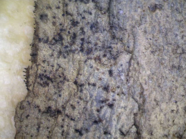
With these strange objects, the same cannot really be said. The above photo is the home of these weirdos. What can you see in this distant photo of the wood. There is a white/grayish mold around the edges. And, if you took liberties, you might say that the edges are also, ever so slightly, textured?
Let's zoom in and progress forward a bit in time.
This is a few days later, and magnified about 40x with a hand lens.
Around the wood, the mold has been disappearing, except for this corner where it still clings to the surface. We an see now that the mold consists of individual strands. These are, more than likely, hyphae, or asexual structures that grow and conglomerate together to form the sorts of moldy fuzzes you sometimes see on food or plants.
Let's zoom way in.
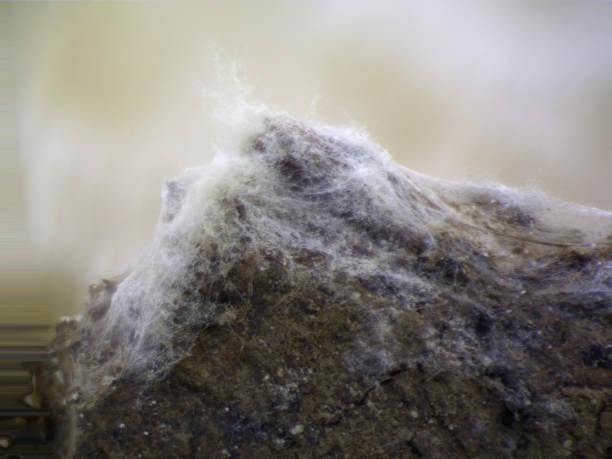
At roughly 80x magnification, the corner of the threadlike mold takes on the appearance almost of a snow capped mountain.
This photo is telling in a number of ways. First, in terms of the characteristics of this fuzz, we can now be certain it consists of interwoven, filamentous strands of white hyphae. It looks almost like a mycelium of sorts - and in a sense it is. More than likely, the final blackberry structures are directly related to this white fuzz - and were formed by it in the same way that a mushroom cap spawns out of a mass of mycelium.
If you look up at the first photo of wood, on the edges, you can see they are textured, in addition to a little fuzzy. I believe those textured areas are the developing sporangia, or spore bearing masses which I have visualized here. However I was not looking closely enough at the time to investigate because I simply wasn't looking for something so tiny. Slime molds are much bigger, in general, than these little guys.
This photo is also relevant to the macro-photographic process in general. If you look on the far bottom left of the photo, you can see an area of distortion. I have kept this in demonstratively. The way this, and the other photos you see here work, is I take anywhere from 5-15 photos of a subject. In each photo a small slice of the subject is in focus. Then I run all the photos through an awesome piece of software called Zerene Stacker. The program identifies the in focus areas, aligns the various photos, and then stacks them on top of each other. The result is a photo that is entirely in focus, albeit with some visual distortions resulting from the cumulative realignment of each photo.
Because the objects being photographed are so minuscule, the really difficult part is finding them at the most useful angle and lighting them appropriately. Thankfully the modularity of the microscope I bought allows for a great flexibility in both these regards. Nonetheless, it is fair to say that the photographic process is now taking as long to produce, in and of itself, as many of my older posts took in their entirety.
But the result has been fruitful, if perplexing. As I was scanning the surface of these hyphae at even greater magnification, I saw something that blew my mind. Infinitesimal, living, moving, eating (?) creatures!
These gorgeous, shiny and translucent bugs were roaming all over the white fuzz of the mold, and you can see them appear to either get stuck in the tendrils, or slowly attempt to ingest them. It's totally unclear. You can also see, if you look very carefully, that the bugs have bits of the hyphae attached to them as they walk around gingerly over its surface.
But the most amazing part of this, from my perspective, was at the roughly 2 minute mark when a second bug enters the frame and we watch it apparently eating the hyphae. Most remarkable are the three or four black dots stuck to the bugs rear end.
These are the spore bearing sacs which fall from the blackberries! They adhere to the bug and it carries them around to aid in spore dispersal!
Something about that single piece of information drove home most concretely for me that this tiny piece of wood bears its own flourishing ecosystem. There are multiple life cycles playing out inside of this petri dish, with at least two disparate organisms surviving interconnectedly, one with the other.
Let's take a closer look at the real star of the show.
This photo is zoomed in about 60x, more than my hand lens would allow.
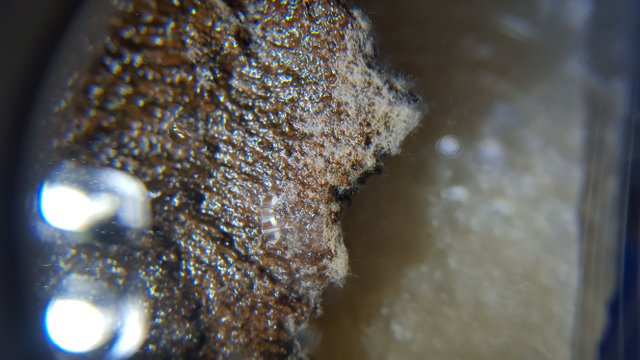
At this point, most of the white filaments had disappeared completed, and in there place were left these small black dots. It was the discovery of these small stains, and micro structures that first spurred me to get the dissecting scope. I was about to throw this piece of bark out as a failure, when I noticed structures so small they were barely visible with my hand lens.
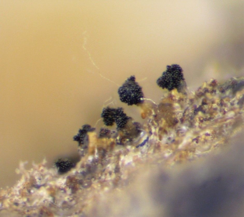
The structures still have the remaining filaments sticking out of them.
Each of these sporangia are sub-millimeter in size, in all dimensions. That would make the individual black sacs absolutely tiny. Nonetheless, I was still able to visualize them pretty well - at least well enough to get a clear sense of the sporangia's general structure.
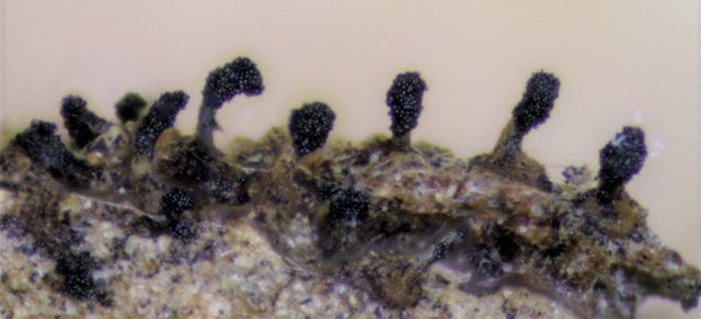
The stem portion appears to be yellowish in some of the pitctures, as here, and the sacs black in color. You can see why I refer to these as blackberries.
Let's take a closer look at the individual fruiting bodies.
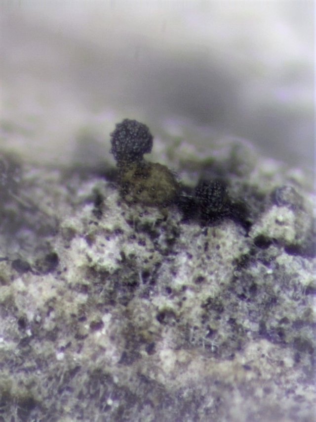
The structure on the left has a rudimentary stem and is somewhat elevated, while the structure on the right is sessile and has no stalk.
Generally, the fruiting bodies varied in this characteristic based on where they were placed on the piece of wood. The structures growing on the sides of the wood, perpendicular to the floor, had stalks, while the structures growing on top of the wood did not.
this photo also give some insight into the dispersal process of the black orbs. You can see how both structures have begun shedding the black orbs. I don't know whether the beetle creatures we saw before eat these spores or simply pick them up in passing.
Here's a sessile grouping we can look at in more detail.
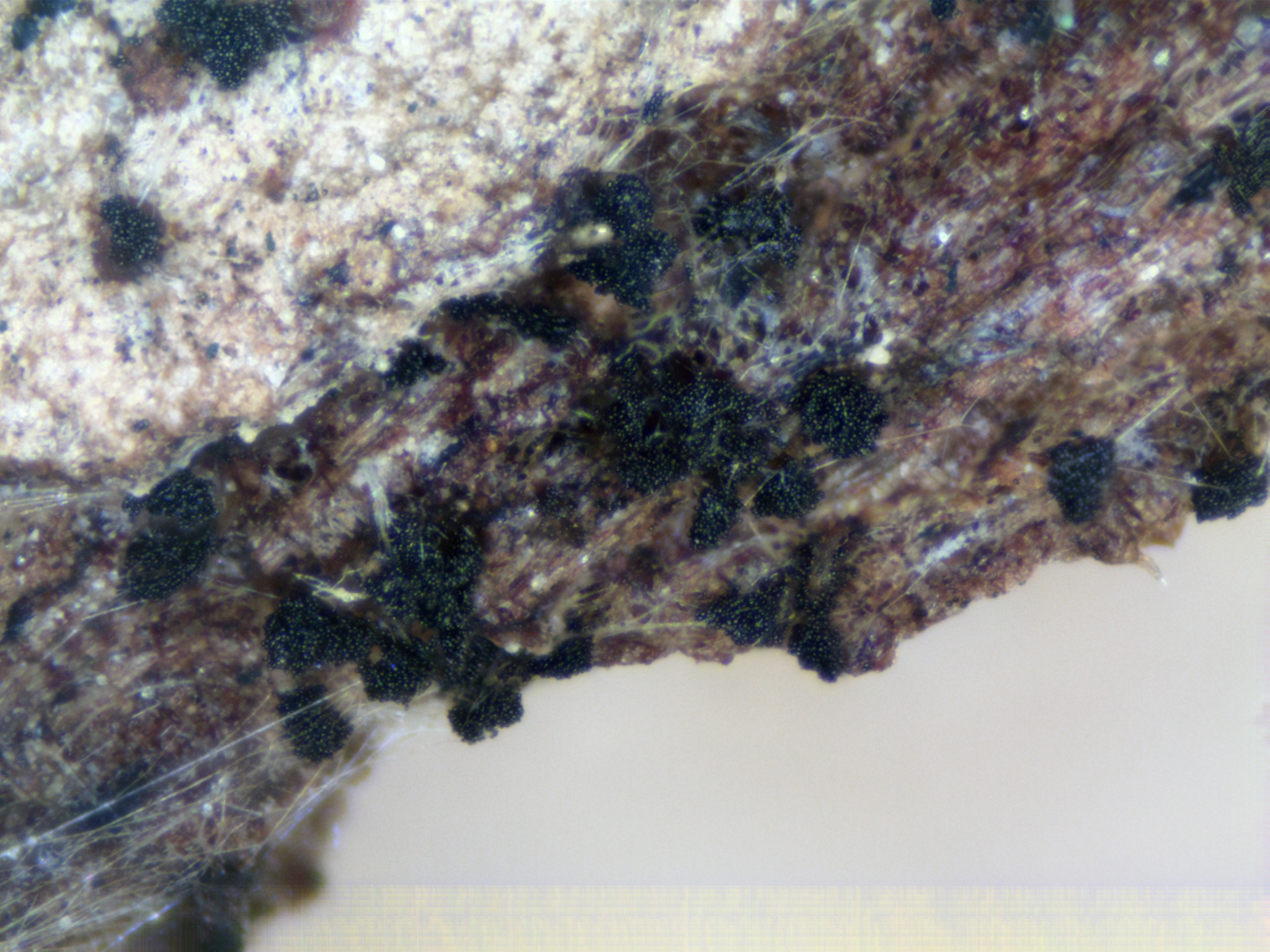
This group better highlights the interconnection between these spore bearing structures and the original hyphae fuzz.
You can clearly see the remnants of the interwoven strands interspersed among the structures. You can see also just ho prolifically these guys propagate. There is no shortage of them, and more than likely each individual black sac contains multiple spores, just because they seem too large to be individual spores themselves.
However, this is not totally implausible either, and it leads us to the ultimate question here, what the hell are we looking at. My first instinct, itself the result of a certain observational bias, was that they must be slime molds. One possibility that came up repeatedly was Polycephalum Physarum, a slime mold which creates globular looking, blackberryish sporangia in maturity.
However, this notion makes no sense. First, the scale of these fruiting bodies is totally off. They are WAY to small to be P.physarum. But, more importantly, this idea simply ignores the fungal mold-like hyphae that both covered the entire piece of wood for a time, and which can still be seen interspersed with the fruiting bodies.
Slime molds do not develop from hyphae. They develop from an amoeba-like state, into a plasmodium that can move around, slime-like, and finally maturing into a spore bearing body called a sporangia. Because these things began life as a fuzzy mold like substance, slime molds are basically out the window.
A user on reddit was the first to inform me that even rudimentary fungal molds can progress beyond simply fuzz and develop fruiting bodies. Further reading as presented to prime, if broad, candidates for where this particular organism falls in the scheme of things.
One possibility is that it is a form of zygomycota. These are most frequently associated with sugary substrates, like strawberries. However, it seems unlikely that these are zygomycotes, because most of those species are known for their singly spore bearing strands in maturity, whereas these guys had a very different final structure.
Which leads me to think these are one of the multitudes of ascomycota molds. The Ascomycota fungi represent their own division in the kingdom of Fungi, and represent the majority of known species. These fungi are primarily distinguished, morphologically, by the microscopic presence of spore bearing structures called asci - derived from the greek word for a "sac". Spores are formed within these asci and then, by some means or another, eventually dispersed.
We've only really covered one ascomycete species here, and that was Cordyceps capitata. Whether this organism also falls into the division might be illuminated through microscopy.
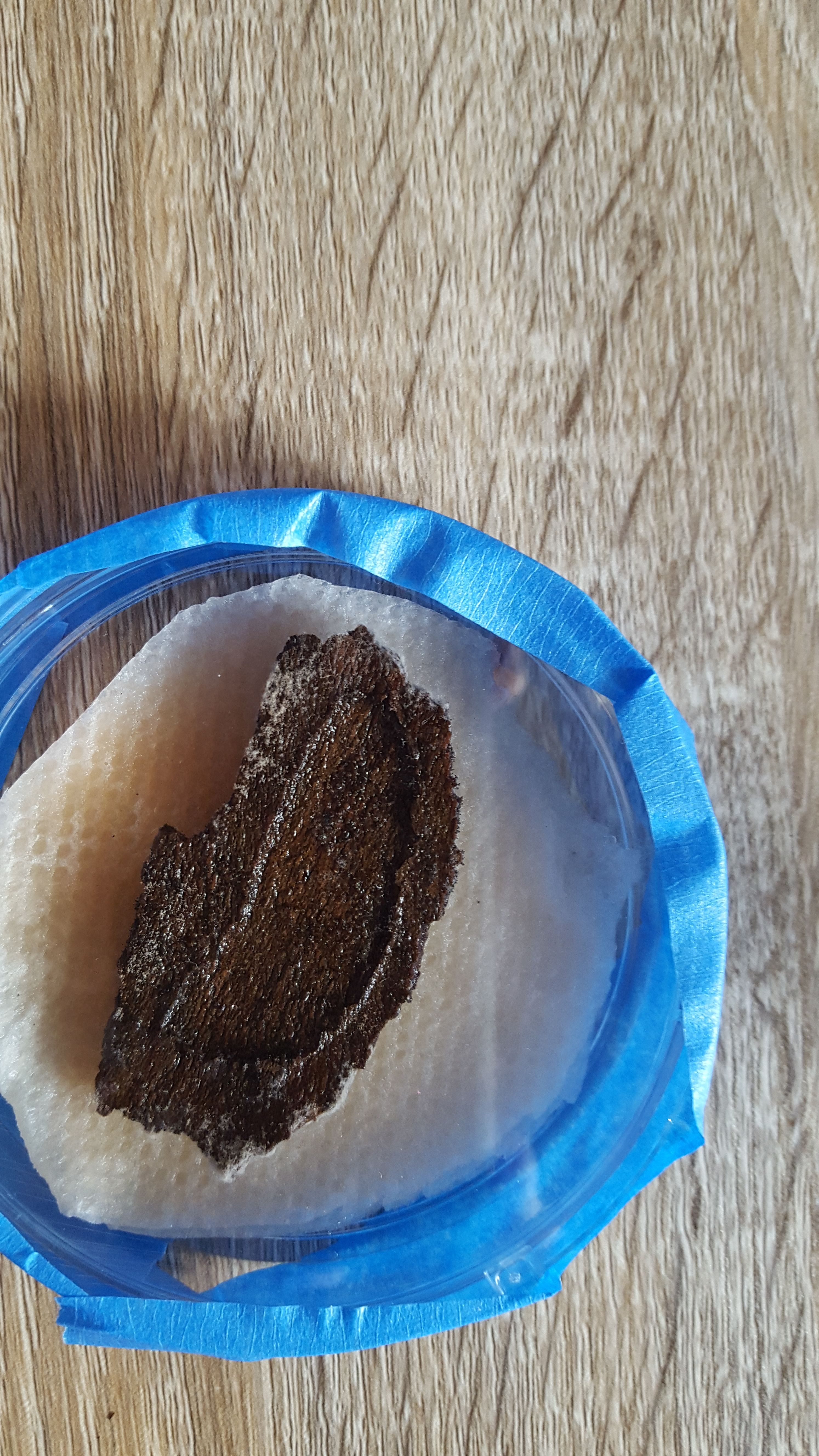
In order to get a sample mounted on a slide, I had to stumble around like an idiot with a dissecting needle. The process is sort of interesting, if a little frustrating to watch.
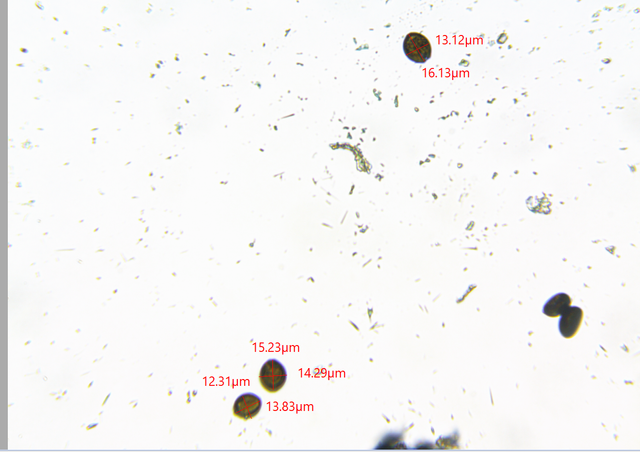
Under the microscope, the small black globes can be measured.
They are on the large size, for sure. So large that I felt fairly certain they were not individual spores, but spore bearing sacs, or themselves essentially macroscopically visible asci.
One microscopic photo in particular convinces me.
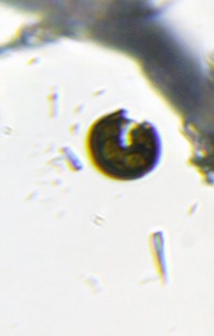
Here we can see what appears to be one of the sacs splitting open!
Moreover, you can also see the hint of structures inside of the circle, both behind the outer skin and poking out from within it.
This image supports the idea that these black orbs are spore bearing sacs, which subsequently break open and release their spore mass. It also supports the idea that I need a better compound microscope. With a 100x oil immersion lens I would be able to tell SO MUCH MORE about these spores. With that in mind, I will be upgrading sooner rather than later.
Where that leaves us with this organism is, likely, in the division of Ascomycota, but not with much more clarity. None of the resources I was able to access was able to provide clear answers. As a result, I can't draw many other conclusions about these things. On the one hand, this is frustrating - but on the other hand, it's kind of awesome. The lack of readily available information about micro-fauna like this means that there is ample opportunity for discovery. As the NYMS gets involved in DNA sequencing with the help of amateurs, I aim to collect and preserve these kinds of samples. There may not be much chance of me discovering a unique species of large fungus, but the chances are much better at this scale!
Photos Are My Own
Microscopic photos were taken using an AmScope SM-4TZ-144A dissecting microscope and an AMScope M150B entry level microscope. The camera lens is also AMScope, MU300.
Information Sources
The bulk of the information in this case was derived from a multi-part posting of these photos and the circumstances of their being taken on several forums involving the study of Fungi and Slime Molds. The information was shared on The Slime Mold Collective, The NYMS Facebook page, and the subreddits /r/mycology and /r/slimemolds. Users from the slime mold collective and reddit both expressed doubt that this was a slime mold and was in fact a fungal mold.
Efforts were made to identify the species using the following books:
[1] Myxomycetes A Hand Book of Slime Molds By Steven L Stephenson and Henry Stempen
[2]The Audubon Society Field Guide To North American Mushrooms by Gary Lincoff
[3]Mushrooms Demystified By David Arora
[4]And, at the suggestion of members of the NYMS, "The Kingdom of Fungi", by Jens H Peterson, which I purchased especially in response to the quandary of this and other small species. It has not revealed precise species, but has shed light on these and other questions. It is one of the most beautiful books I've ever owned, and I can't suggest strongly enough that you all purchase it.
[5]Zygomycota
[6]Inaturalist was a resource, but provided no concrete answer
[7][A big thanks to the various members of the Slime Mold Collective, NYMS and /r/mycology and /r/slimemolds for their input. The cumulative comments helped provide a modicum of direction where I had none originally.
Wow, wow, wow, you have gone to such detail, not only in your pictures but also your post! You have a new follower and I'll be checking your back catalogue too!
I do love mushrooms, I would love to be able to forage properly but without proper guidance I guess you are playing Russian roulette! Something I am not willing to do. Your previous posts will enlighten me somewhat. Could you recommend any books on the subject?
Also being Vegan I have eaten my fair share of Fusarium venenatum.
Thanks for the information. A real eye opener.
I'm glad you're so interested -in terms of recommended reading the books listed in the information section of this post are a great start - in particular the Kingdom of Fungi is great as it takes a global view.
Otherwise it depends on where you are in the world. But rest assured, wherever you are there are definitely mycological resources tailored to your general area. My guides are primarily tailored to the North East of North American cause that's where I do most of my searching.
But come April I'll be joining a group down in Chile and that will require inquiry with an entirely different set of resources.
And btw, I too don't eat my finds. I think the risk/reward ratio is all out of whack, even if you can ID very well. Growing mushrooms for food is a different matter however and we'll be addressing that in the next few weeks as well.
Brilliant thanks. I am in Ireland so will have a look at the kingdom of funghi book. I'll be looking forward to your post on growing for food.
Thanks again and keep up the great work.
Thank you for writing such an informative thorugh post. I have become very interested in all things 'mushroom' since i heard about Korean Natural Farming a little while ago. i have also been reading about how the bees are dependent on the network of fungi in the soil and on plants. I am hoping to get the book how mushroooms can save the world,this is a fascinating subject!!!
Great article, i loved the passion with which you described your discovery and the pictures are beautiful. Long live participative science !
As always, very complex and intresting. Keep going, great job @dber :)
Well @dber. I can see your new microscope is heavily in use. I am very happy for you!
It is - and Thursday will be even cooler insofar nature will prove itself to be significantly more lit
to me very Interesting photos,about these organisms I yet never heard reading pleased thank you for information.A good post likes me
Very intesting @dber .. I learn new things from you on how to make good post about #steemstem , thanks bro ;)
World of Photography Beta V1.0
>Learn more here<
Thank you for participating in #macrophotography, the weekly selection will be released on Friday.
You have earned 5.30 XP for sharing your photo!
Daily photos: 1/2
Daily comments: 0/5
Multiplier: 1.06
Server time: 15:36:36
Total XP: 45.35/100.00
Total Photos: 8
Total comments: 4
Total contest wins: 0
Follow: @photocontests
Join the Discord channel: click!
Play and win SBD: @fairlotto
Developed and sponsored by: @juliank
strange organisms.
And I in shock for not how many hours for you already 185 dollar and for me even a 1 dollar not when was not it in what secret?