The Amateur Mycologist #31 - Metatrichia vesparium - Behold Some Serious Slime Mold Weirdness
These posts are not for foraging. They are intended for entertainment and intellectual satisfaction only. These posts are not a field guide nor comprehensive in any way - their accuracy is not assured in any way. Do not eat wild mushrooms unless you are a professional, have substantial professional assistance or have a wealth of personal experience with a specific species. Do not make any foraging decisions based on these posts. To do so could be dangerous or life threatening.
This post does not include edibility or toxicity information
A NYMS member has taken the time to consult a fairly obscure field guide on North American Slime molds by Thomas Houston. And, I'm glad to say it makes reference to m.vesparium having SUBGLOBOSE spores! With that characteristic clarified I now feel confident enough in my ID to upgrade this to a full species post. I will do the other edits later tonight!
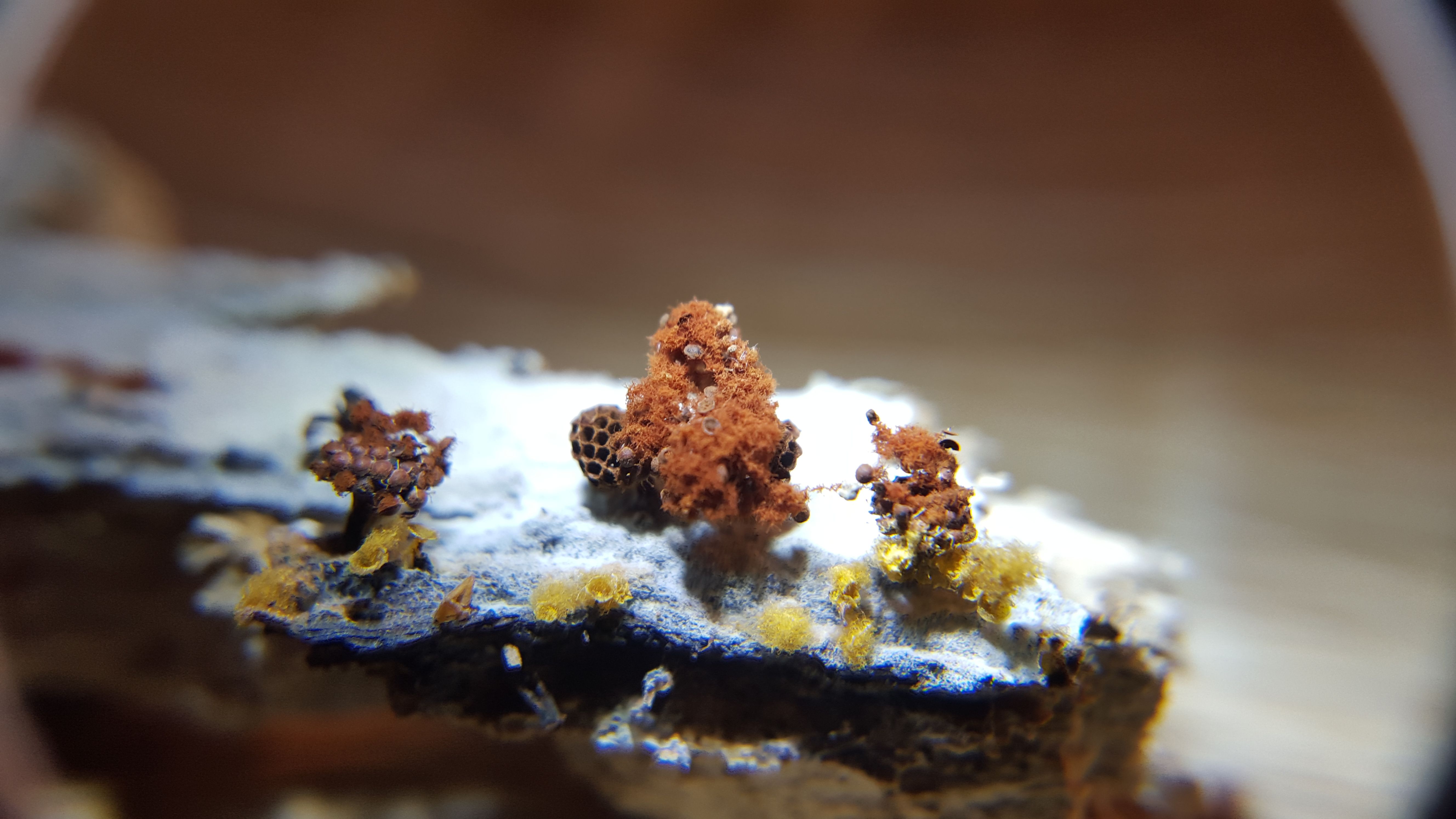
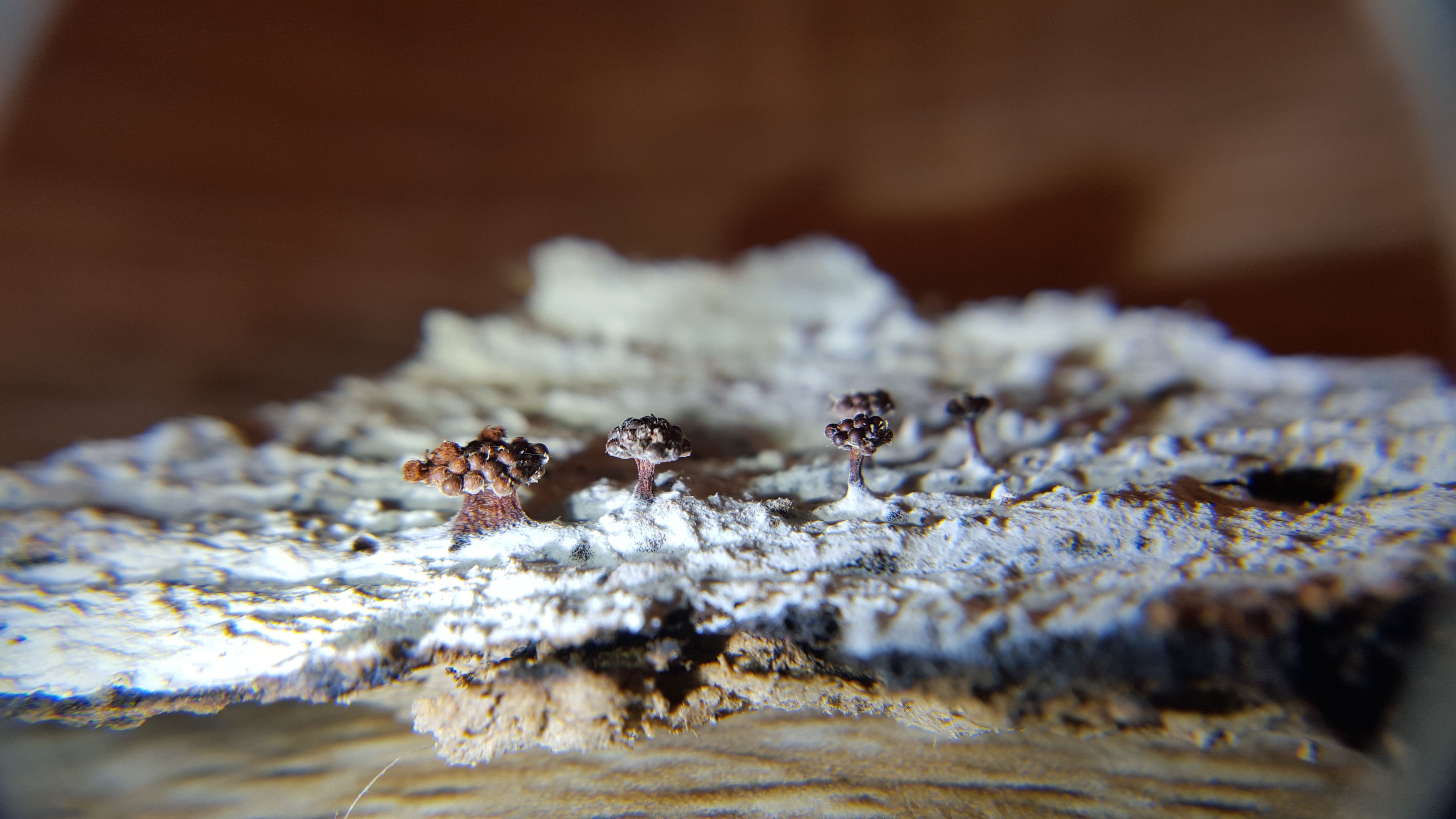
What is this bizarre panorama of chaos?
There's a lot going on in this photo and we're going to need to tease it all apart. But initially, we can summarize by saying it contains two different species of plasmodial/acellular slime molds, or Myxomycetes. There is the predominating red sporangia - or spore containing structures - on top of stalks and covered in a red fluff called capillitium, which is interspersed with spores.
Then on the bottom of the small ecosystem of wood are several yellow patches. These are a different species, but you can make out to the left of the photo some kind of sporangium structure, as well as bright yellow capillitium on the far right.
Let's label this same picture and take another look.
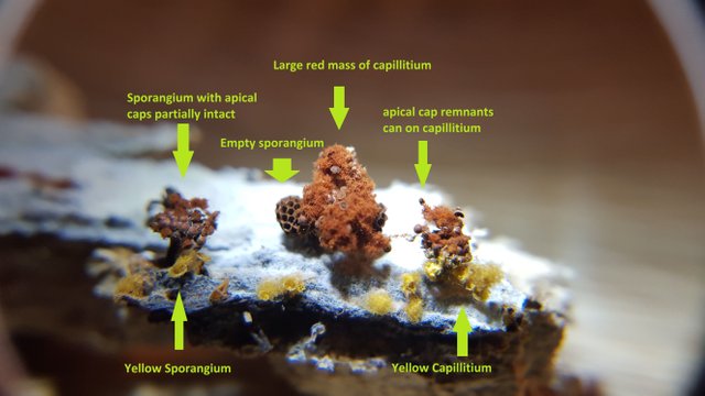
Hopefully that clarifies the various elements in the photo.
Today's post is going to be about the top, red, stalked organism, while next Tuesday's post will be an effort to decode the yellow one.
Now, let's try to get a look at the same scene without magnification in the wild.
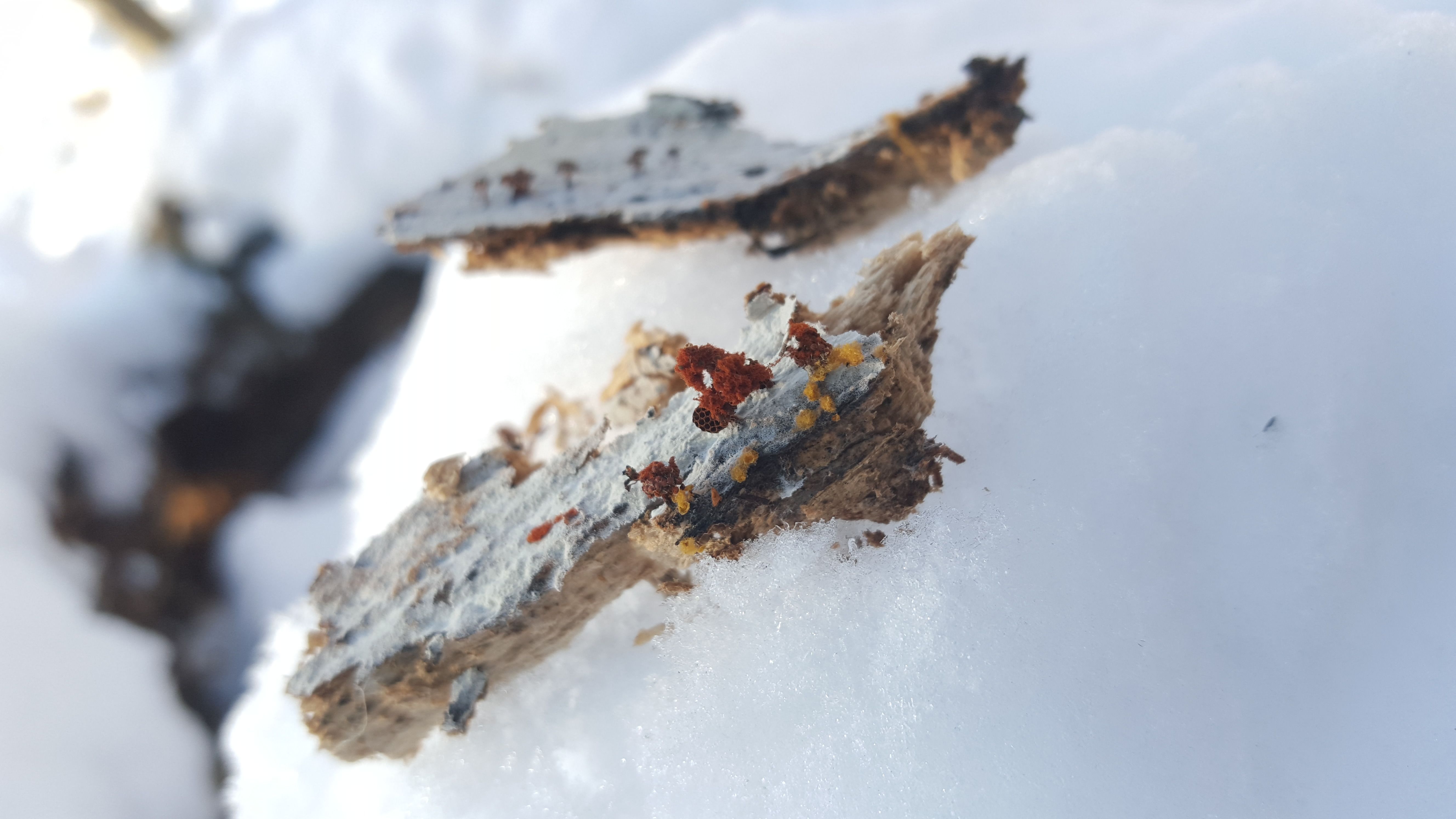
The explosion of chaos in the magnified picture above occurs on a tiny scale.
These organisms - the final sporangia of these slime molds - are quite small and, sometimes, difficult to see when foraging. Other times, they amass in impressively sized colonies - either as the final spore bearing structures or as a pre-sporangia mass of slime - at which point they can be impossible to miss.
This was one of the former cases - and the only reason I found these tiny guys is because it was cold and snowy - and failing to find big, flashy mushrooms, I decided to zoom my focus in to a smaller scale. So if you ever take a walk in the forest and see someone meticulously analyzing every inch of a decomposing log, you may now have a sense of what they're searching for.
Let's start breaking this species down by characteristics.
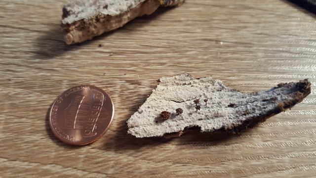
This shot gives an even better notion of the scale involved.
These sporangia are not much more than one millimeter or so in their dimensions, at the largest. It should be noted here that there is actually a third organism likely involved in this piece of wood - and that is the white background. More than likely these sporangia are the result of a sporulation from some time ago. However the sporangia nonetheless survive structurally for awhile afterwards. The white floor they are growing from is the result of some kind of organism - I just don't know what.
It could be the desiccated remains of the Metatrichia plasmodium - although I don't believe that is white in color. Or it could be the sclerotium of a different slime mold - which is to say the long term hibernation mode some slime molds use to survive harsh environmental conditions.
Truthfully, I don't know what the white stuff is - and I wasn't able to discern much under the microscope. But luckily, these M.vesparium did not disappoint.
Here you can see the full sporangia structure, held aloft on a sometimes quite thick stalk.
These were not more than 2mm high at the maximum. These particular sporangia have not yet burst and released their fluffy red capillitium. Instead, the "goblets" which contain the spore mass here remain whole, and the apical - that is "relating to or denoting an apex" - cap is still firmly attached to the goblets.
The photos above depict what happens when the sporangia are ready to release their spores. The goblets break open by the coiled force of the capillitium, which springs out in a big scrunchy mass.
Remember, as we go through these photos that we are looking at the final stages in the life cycle of an organism that is profoundly different from a fungus. Even though these structures appear convincingly fungal in nature, the rest of the slime mold's life cycle is nothing like a fungus at all.
Let's get the spore mass onto a slide.

Using a razor blade I was able to extricate a hunk of the capillitium.
I cut this big piece a bit with the blade after this photo was taken in order to get a smaller piece to look at. I also sort of blotted the slide with the whole mass of capillitium in order to get it to drop a layer of spores. I placed one drop of distilled water on the spores and the mass of capillitium and placed two different glass slide caps on each sample, on the same slide.
Let's see what we get.
Ah huh...
I guess I should have suspected this kind of structure, given the springiness of the capillitium, but still seeing it magnified was a surprise.
ENHANCE
How strange is that?
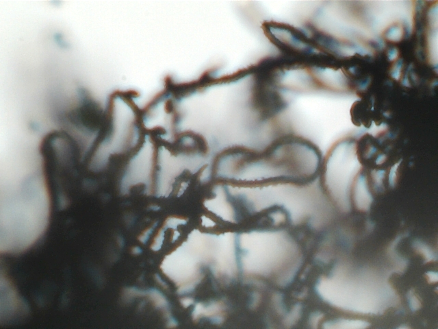
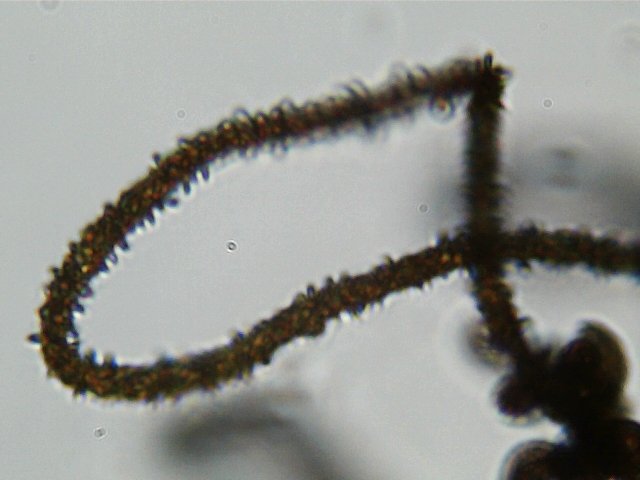
Here, at the microscopic level, things reach a new degree of confusion. This is a roughly 400x magnification of a single strand of capillitium. Although these strands form inside the same cavity in which the spores are produced, their production has nothing to do with the production of spores. The spores and the capillitium develop concurrently in the sporangia, but the capillitium is entirely sterile.
However, the capillitium do serve a spore related purpose, by all accounts. Their springy nature breaks open the sporangia to release the spores, and the dense, hairy texture can cause spores to stick to the capillitium for a time so as to better disperse from inside the sporangia.
Although I am fairly satisfied with this first microscopic slime mold observation - it also creates some problems. I believe the combined failure of proper sample preparation, with the lower quality of my camera and scope, makes capturing the fine details of the capillitium nigh on impossible.
For instance, these capillitium should be composed of 3 to 5 spiral bands - however that is simply not clear one way or another in this photo. The Handbook does specify that the individual coils of capillitium do bend 180 degrees about half way down their length - and we can see in this photo, as well as in the photo at 100x magnification. We can also see the spines running down its length. I tried to measure them, but I got mixed results that were not, I think, particularly edifying.
Unfortunately, the microscopic photos of the spores only added to the confusion.
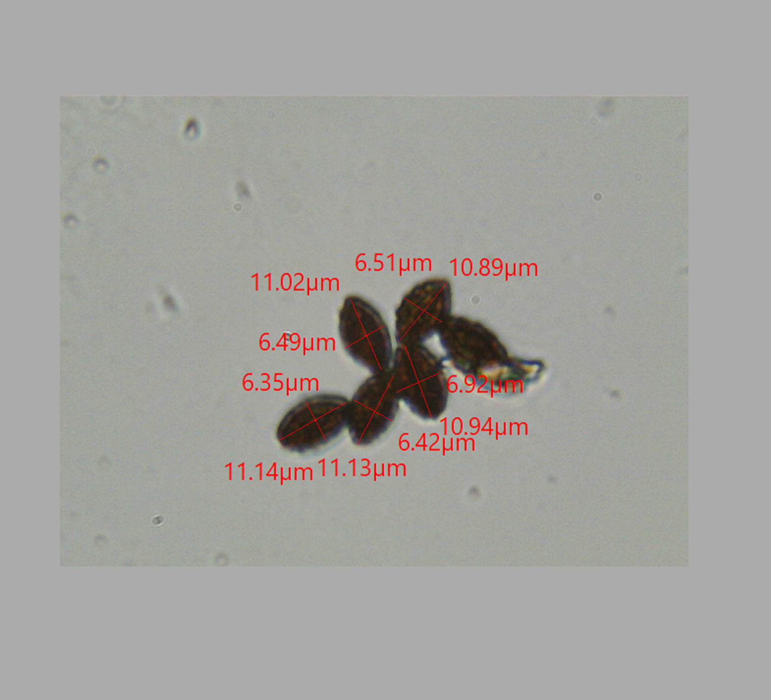
These robust, ellipsoid looking spores further confuse matters.
Both of my sources describe the spores of this species as round, but here we clearly have a different shape. The length of the spores matches the range given in the Handbook for the diameter, wherein it is described as 9-11 microns. It also seems to me you can make out the "minutely warted" detail as well in this photo. The color, however, seems quite dark - although I did not prep this with KOH or a stain, and so they may be less translucent.
The question is, why is the shape so different? I don't know! There's so much I don't know about slime molds. This fruitful, tiny piece of rotting wood was only the second slime mold source I've ever found.
I was very tempted to make this into a full on species post for M.vesparium. This temptation was based on the obvious macro-characteristics of the sporangia and capillitium, which match the Audobon guide and online pictures precisely.
However, I have said many times before that if you are not 100% certain about the identity of a mushroom - or in this case a myxomycete - then you don't really know what that organism is - and to act like you do, however much you may want the organism to be what you think it ought to be - is foolish.
The fact is - although these specimens fit the macroscopic bill almost perfectly for M.vesparium, the spores establish a substantial distinction, and one which I can't simply ignore. But, whatever the heck this thing is, it is splendiferous and makes for some awesome macro photos. Next week we'll take a closer look at its bright yellow cousin.
THIS POST IS NOT INTENDED FOR FORAGING PURPOSES AND TO USE IT FOR THOSE PURPOSES WOULD BE DANGEROUS. DO NOT HUNT WILD MUSHROOMS WITHOUT RELYING ON A COMBINATION OF PROFESSIONAL FIELD GUIDES, IN PERSON PROFESSIONAL GUIDANCE, OR IN PERSON GUIDANCE BY SOMEONE TRUSTWORTHY WHO HAS COPIOUS LOCAL, SPECIALIZED MUSHROOM HUNTING EXPERIENCE. FAILURE TO DO SO CAN RESULT IN GRIEVOUS PERSONAL HARM OR DEATH.
Photos Are My Own
Microscopic photos were taken using an AMScope M150B entry level microscope. If you use microscopes, I'm quite sure you've never heard of this model - but its cheap and available on Amazon. The camera lens is also AMScope, MD35 - by far their crappiest microscope camera. But, still capable of material, relevant, and in some cases, dispositive, data collection. Lastly it should be noted that the precise magnifications are not easily deduced using the camera - but based on relative spore sizes compared to known microscopic photos from Kuo and other sources, I estimate 40, 100 and 400x.
Information Sources
[1][Gary Lincoff, Audobon Society North American Mushroom Field Guide, Cordyceps capitata, p.852]
[2][Myxomycetes A Handbook of Slime Molds - Steven Stephenson and Henty Stempen, p.137-138]
It looks like a cake :) Okay, I should sleep more :p
More seriously, I was wondering, when you don't know some stuff, is there any place where you could go and ask for the question?
Several - i just joined the Slime Mold Collective - facebook has the mycological society of ny - and reddit is also forthcoming. Often the gaps in my knowledge turn into edits subsequent to posting - thats what happened last two posts actually - tworeddit users who know a ton about slime molds told me where i was wrong and that resulted in a ton of edits.
I expect a similar thing to happen here in the next couple of days :)
Having some comment issues - sorry - i meant to add that these other resources are available when the books and online resources fail to completely edify.
Case in point - I referenced this to members of the NYMS and one of them looked up the species in an obscure book on North american slime molds which confirmed that the spores of this species have been seen to be Subglobose. This clarifies my final big hesitation about the species and confirms my ID. So i'm going to reedit this whole post to make it into a species post and add the new information source.
Similarly the white stuff is likely to be a form of resupinate fungi - which came after the slime mold sporulated. Also a tidbit from a NYMS member that bears out as a good hypothesis.
Another excellent review! Thank you.
Thanks for taking a look, as always - I know the last few posts have moved into pretty non-practical subject matter - next thursday I'll be back to a more conventional fungal post about sulfur tufts and the dangers of mis-id'ing close species on the same growth medium.
Thanks again for sharing your wonderful learning with us. As always it is very impressive, def inspiring me to get a microscope. How is your experiment/ growing slime coming along?
You definitely should get one - so many things to see at the mircoscopic level.
I need to post an update - so far it's just limited to that white fluffly stuff - but its still quite moist so we'll see if it progresses in the next week or so.
beautiful photography and what an interesting post with great insight!
Wow, fantastic photography, info and insights. You must have studied a long time to become so informed.
Thank you for sayong that - but this whole blog is really a form of journaling about my learning experiences. When i find a new species I try to become as informed as possible about it - but the extent of my general knowledge is only in its infancy. Plus, although I make efforts to avoid mistakes, they definitely happen all the time. I try to fix such issues when they arise by cross referencing my posts with other specialized communities who enjoy the subject matter.
Well, I have to say I was very impressed and I have always wanted to do some foraging but was unsure of so many berries and shrooms that I get nervous and stick to the grocery store or grow it myself. I am an avid grower for over 45 years and love it. Love to take care of my family in an old fashioned manner.
Actuakly, I'm the same way - I almost never eat my foraged finds. Even as I become more knowledgable i still can't bring myself to eat them. That's why my disclaimers are so huge and why I generally dont talk about edibility. I like to learn about these things - but outside of emergency i'll stick to more conventional foodstuffs.
I so agree and don't blame you on that decision.
amazing post and info !
Thanks
like your post.thanks for sharing
beautiful photography & very nice post. i like post & resteem this post
Hello Friends, How Are You
Thank You Friend nice To Meet You
the Best And amazing.
Nice To Meet You All #steemians
My Name Ary, Account name @roygarudakirana I Am Coming From Aceh Sumatra National Liberation Front (ASNLF)
I Say Welcome And Happy To You Here #steemitor#eSteem.
Vote @good-karma As Our Witness.
Do not Forget Follow Me Also @
I would like to express my gratitude.
nice work. upvoted and followed.
this is my blog(please upvote and follow if you like):
https://steemit.com/life/@benainouna/10-reasons-why-plastic-bags-should-be-banned