The Amateur Mycologist - Random Find - Best Guess Is A Hemitrichia or Trichia Species Based On Rudimentary Microscopy
These posts are not for foraging. They are intended for entertainment and intellectual satisfaction only. These posts are not a field guide nor comprehensive in any way - their accuracy is not assured in any way. Do not eat wild mushrooms unless you are a professional, have substantial professional assistance or have a wealth of personal experience with a specific species. Do not make any foraging decisions based on these posts. To do so could be dangerous or life threatening.
This post does not include edibility or toxicity information

Last time we considered the red portion of this picture - now we move on to a far more difficult puzzle piece
What is that yellow stuff? No doubt at all that it is the final part of a slime mold's life cycle, but in terms of what slime mold, I have more questions than I do answers. Spoiler alert - we are not going to get very far in ID'ing these yellow guys, in part because of microscopy issues that the new camera may rectify.
But first let's take a closer look at that photo.

Compared to the downright gigantic structures of M.vesparium, these yellow guys are tiny.
Tiny and difficult to visualize well enough to get a sense of the structure of their fruiting bodies. Or at least, what remains of the fruiting bodies.
The clearest indication of what the fruiting bodies might once have looked like, in part, is on the far left of the above photo. Keep in mind, this photo was taken with a 40x hand lens, and the fruiting body of this yellow species is so tiny that it is still hardly visible in any detail. In order to see structures like this, I'm going to need a dissecting microscope - which is currently third on my list of future equipment purchases.
Still, we can come to some conclusions about these yellow guys using what equipment I already have.
Let's dive into the microscope and take a look at that yellow fuzz.

First thing, we can see the capillitium en masse. This is an important observation to get us into the Trichia/Hemitrichia genuses. Let's zoom in.

Here we run into the problem of a a lesser microscope.
This looks to be what is called an "elater" - or a single free strand of capillitium that appears in many species in the Trichia genus. However, in order to identify the specific species, we need to be able to take a closer look at the elaters. One thing we can see here and in the photo right above is that the ends of some of the elators appears to be pointed. We can also see that there is a distinct texture to the elaters.
However, we cannot tell based on these photos precisely how many strands make up the elater's texture - which could be anywhere from 3 to 5 individual spirals. Here we can only see the hint of a woven texture. Plus, I messed up and forgot to measure the elaters when I originally took these photos - and now, because the resolution differs from my viewing software's calibration, I cannot reverse engineer a measurement.
Things do not get clearer with the "spores"
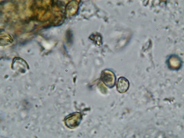
Is that them there?

How about there attached to an elater?
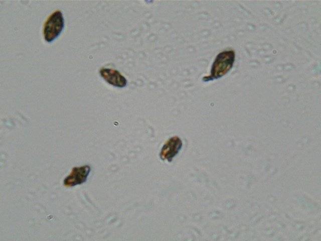
Or could these be them instead?
My initial foray into slime molds really highlights the need for an oil immersion microscope with a greater degree of magnification. Here, I have a sample that, for one reason or another, has not presented me with an image unequivocal enough for me to discern which objects are even the spores, let alone allowing me to try to make an identifying judgment call.
In this case, either because of the nature of the sample or my preparation, I botched it. I can't make out with any certainty which part of these final images are even relevant. And this a major problem, especially when it comes to myxomycetes.
Even more than many mushroom species, myxomycete identification is often based on microscopic characteristics - especially if you intend on getting down to a species with any degree of accuracy. Some species have such obvious, even explosive macrocharacteristics, like M. vesparium, that microscopy is mostly confirmatory.
But in the case of these yellow guys, found at this point in their life cycle, microscopy is not just a confirmatory procedure - it is absolutely central to achieving an ID. Based on the combination of bright yellow macro-characteristics, combined with the presence of elater based, yellow colored, and woven textured capillitium, I think it is likely that these guys were in the genus Trichia or Hemitrichia. But, beyond that, I daren't even fathom a guess.

Although this ID process was not so successful, the quest to further explore slime molds continues with my moist chamber experiment.
After almost two weeks, the initial chamber is still quite moist, but visible growth is still limited to a white fuzz around the edges. However, I had to disturb the sample by moving it from a household tupperware into one of the new empty petri dishes I purchased on amazon. It is generally frowned upon to open the chamber before sporulation, but my wife wanted a container to put her salad dressing in.
I am not super optimistic about this first piece of bark at this point. However, far from giving up, I've doubled down.
Two days ago I was in Staten Island and harvested several new bark samples

I will be dividing these up into bite sized pieces and setting up several new moist chambers simultaneously. Between all of them, we should get something over the coming weeks.
I'll document that moist chamber process, along with a refresher on microscope calibration for my new camera, next week.
THIS POST IS NOT INTENDED FOR FORAGING PURPOSES AND TO USE IT FOR THOSE PURPOSES WOULD BE DANGEROUS. DO NOT HUNT WILD MUSHROOMS WITHOUT RELYING ON A COMBINATION OF PROFESSIONAL FIELD GUIDES, IN PERSON PROFESSIONAL GUIDANCE, OR IN PERSON GUIDANCE BY SOMEONE TRUSTWORTHY WHO HAS COPIOUS LOCAL, SPECIALIZED MUSHROOM HUNTING EXPERIENCE. FAILURE TO DO SO CAN RESULT IN GRIEVOUS PERSONAL HARM OR DEATH.
Photos Are My Own
Microscopic photos were taken using an AMScope M150B entry level microscope. If you use microscopes, I'm quite sure you've never heard of this model - but its cheap and available on Amazon. The camera lens is also AMScope, MD35 - by far their crappiest microscope camera. But, still capable of material, relevant, and in some cases, dispositive, data collection. Lastly it should be noted that the precise magnifications are not easily deduced using the camera - but based on relative spore sizes compared to known microscopic photos from Kuo and other sources, I estimate 40, 100 and 400x.
Information Sources
[1][Gary Lincoff, Audobon Society North American Mushroom Field Guide, Hemitrichia clavata, p.850]
[2][Myxomycetes A Handbook of Slime Molds - Steven Stephenson and Henty Stempen, p.156-157]
I admit that I am having a harder time keeping up as your posts have become so detailed, but thank you regardless for putting this knowledge out there to be found and surely used for years to come!
Sorry! Thanks for sticking in there! I know I said we would do mushrooms again this week - but I forgot it was the holidays - but we will talk about some big ole mushrooms soon enough, I promise!
You are doing a great job. Looking for your next post. Have a great day !
Thank you @azen! Have a good New Year#
As always very informative post, even if you don't identify the type, I'm learning alot about the terminology used and nice to get an update on the slime growing experiment. Have a very happy new year @dber
Happy new year to you and yours @trucklife-family!
I am trying to understand what you posted. I am still reading it, just want to say holiday
Great post! I'm normally quite averse to looking at mushrooms/fungi/mold but I'm sure that they hold many secrets inside so it's awesome that you're sharing your research into these - maybe you'll find the next penicillin!
Also, happy new year!
I should be so lucky!
I'm afraid my research - to the extent what I do amounts to that - is much more rudimentary than all that. But perhaps one day - quite a long time from now - I can start playing with fungi and slime molds as they relate to other organisms. For now, I'm just trying to get my bearing on the fungi themselves :).
Happy new year to you as well!
Interesting blog but there is not detailed