Learn more about the structural characterization of materials through microscopy (atomic force microscope AFM)
Since the invention of optical microscopes, that is, the common microscope, many techniques have been developed to be able to observe smaller objects, the invention of an optical microscope was a scientific breakthrough, but this instrument could only bring objects with visible light through of lenses that approached but not much increase, that is why the scientist as science progressed and more materials were created decided to invent other types of microscopes, since with the optician could not appreciate very small objects, that is, smaller than 1 nanometer, for this reason the man aroused more curiosity to see more inside those objects and where they invented instruments where they could observe objects at visible wavelength, very small scales up to 500 nanometers, and that is where the microscope was invented atomic force (AFM) that does not use visible light to bring objects closer and observe them in an incredible way.
Source
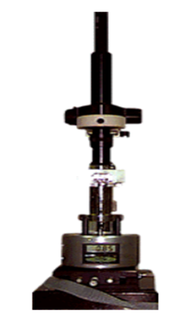
The AFM is one of the first electronic microscopes that were invented, in the mid-80s, its invention was a total revolution for the area of materials and has been used in many fields of science such as chemistry, biology, physics and engineering, since through this type of microscope can be observed surface structures of various materials, and even samples of very hard materials such as ceramics and also very smooth as in the biology to be able to observe cells of plants or any other organism, its versatility and Wide application makes that even today is used.

Current model of the atomic force microscope.
Source
For this reason I would like to talk a little about the AFM and share this post with the whole community.
It is a microscope used to observe surfaces of different objects as mentioned, this technique allows to see and measure structures of different materials and different scales and sizes, in general this technique allows to obtain the surface arrangement of molecules in the field of biology, see material surfaces in the field of materials physics. This mechano-optic instrument has the ability to capture images with nanometric forces on a large scale and that is why today is still the favorite of many.
When we place the object or sample that we want to analyze, this device records the height of the surface of the object by means of a cable or probe, which has as characteristic a pyramid-shaped crystal tip as can be seen in the following figure.
Atomic force microscope.
Source
This probe is connected to a very small cable and in turn very sensitive to detect some force, it has a length of about 200 micrometers in its length although this varies depending on the configuration we want to use.
Basically, this atomic force when detecting when the tip of the crystal approaches the sample, can register a small flexion of the microscopic cable through a laser beam that is reflected on the back side of the same. An electrically coupled system auxiliary displaces the sample of the material analyzed in three-dimensional form, while the tip of the crystal travels the surface of the sample.
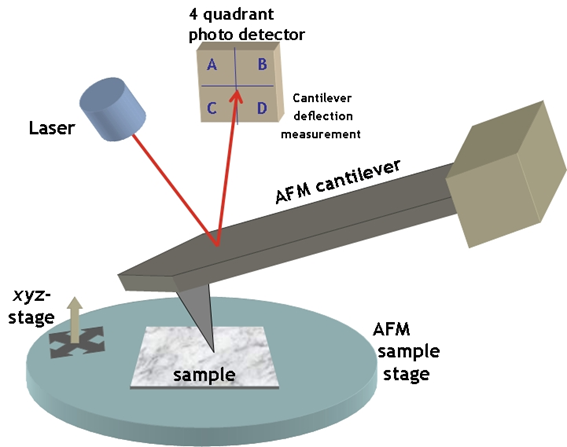
Scheme of the operation of the AFM.
Wiki
The principle of measuring these forces is very simple and it is about measuring the deflection of a spring. If a spring with an elastic constant k is compressed by a force Fz, the compression deltaz of the spring is an indirect measure of the force applied, since by Hooke's law: Fz = k deltaz. The "spring" used for measurements with the AFM, which must be ultrasensitive, is a flexible microlever, microfleje or Cantilever cantilever of elastic constant of the order of 1 to 10nN / nm, and with a sharp tip as shown in Figure.
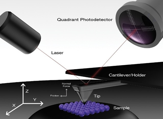
Azonano
Another way of explaining it physically and mathematically can be as follows:
And finally we can appreciate the great resolution of this technique through a computer program that allows to detail in a very amplified all the surface characteristics of the sample, in an impressive way for that time it was something amazing to be able to observe such small objects close to up to a million times.
It depends on three important components that according to the physical characteristics of the material and its properties allow to perform the sweep.
The first is placing the cantilever on the tip of the microscope.
The second one performs a piezoelectric scan that manages the position of its coordinates.
And the third place the feedback loop or control loop.
Now as we know the AFM allows to measure the topography of the sample when in this we slide the tip on the surface as mentioned above.
Another way to operate the AFM is called tapping that allows intermittent contact on the sample by measuring the topography of its surface.
Through this microscope many studies can be carried out that allow to have an important information about the properties of different materials among them are:
The magnetic force that allows to measure the magneticity that the material possesses by means of its gradient, the electric force that likewise allows to measure the force by means of its gradient, its electrical power that exists in the surface of the material, all these by means the physical equation of the gradient.
We can also obtain a phase image that provides us with all the surface information, deformations, oxidation in the area of materials science and bacteria in the area of biology or microbiology.
It also allows us to measure the elasticity of the sample, how soft or hard it can be, likewise the force of friction between the tip and the surface of the sample and many more applications that perhaps we did not have in mind as the Van der forces. Waals, other quantum applications such as the tunneling force, also see the conductive properties of elements or compounds through electrochemical techniques, among others.
Credits:
http://www.usfx.bo/nueva/vicerrectorado/citas/TECNOLOGICAS_20/Topografia/2.pdf
https://nanocienciainforma.wordpress.com/microscopio-de-fuerza-atomica/
http://materias.df.uba.ar/l5a2017v/files/2017/02/Laboratorio-5_Practica-de-Nano-y-AFM.pdf
https://www.bruker.com/es/products/surface-and-dimensional-analysis/atomic-force-microscopes.html
http://www.sld.cu/galerias/pdf/sitios/histologia/el_microscopio_de_fuerza_atomica.pdf
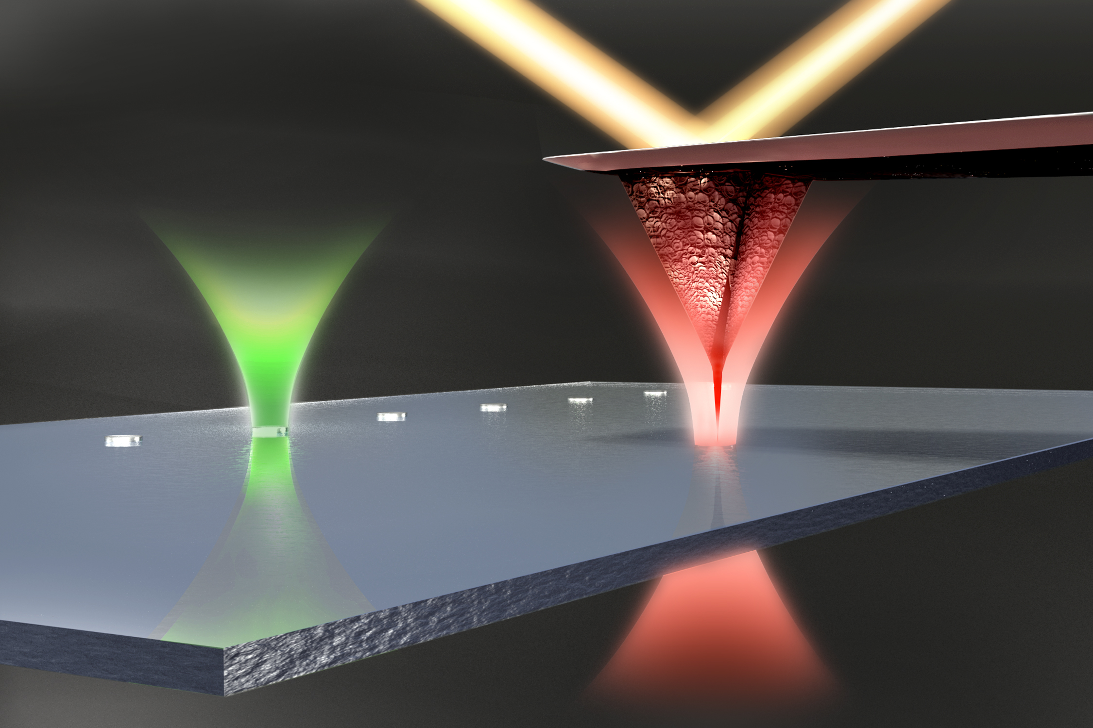
https://jila.colorado.edu/perkins/research/articles/ultrastable-atomic-force-microscopy
https://simple.wikipedia.org/wiki/Atomic_force_microscope
If you want to learn more about microscopy visit my previous publications:
Congratulations @carloserp-2000! You have completed some achievement on Steemit and have been rewarded with new badge(s) :
Click on any badge to view your own Board of Honor on SteemitBoard.
For more information about SteemitBoard, click here
If you no longer want to receive notifications, reply to this comment with the word
STOPThanks @steemiboard