Tools and the Sciences - Exploring the Microscopic World
Among the inhabitants of the microscopic world are bacteria (right) and protists (left). Image Source
In 1976, the Royal Society of London (the avant-garde scientific group of the time) received a letter that would forever change the way we view our world. The author of the letter was Anton van Leeuwenhoek, Dutch upholsterer and amateur scientist. In his letter, van Leeuwenhoek described his surprising observations of a drop of water, where he had seen living creatures he called "animalcules".
What Van Leeuwenhoek had seen were microscopic organisms too small to be seen by the naked eye. Van Leeuwenhoek opened the door to a hidden world through a simple microscope.
Today, after more than three hundred years, modern microscopes have been built with which he would never have dreamed. Furthermore, the exploration of the microscopic world has led us far beyond the "animalcules". However, there is something that remains the same: The door to the microscopic world is still open and there is still much to discover. Maybe one day a letter of yours will amaze the scientific world. Remember, van Leeuwenhoek was not a professional scientist but simply curious.
Since the discovery of Leeuwenhoek in 1676 microscopes have played an important role in scientific research. Microscopes are instruments that produce images, photographs and even videotapes larger than real life (augmented). Most microscopes use light beams to produce the magnification of an object's image. These microscopes are called optical microscopes (optical refers to light).
Optical Microscopes
If you have ever looked at an insect or other object with a magnifying glass, you have used a type of microscope known as a simple microscope. A magnifying glass is just a simple microscope because it only has a lens. A lens is a piece of curved glass. When they pass through the crystal, the rays of light bend. In some types of lenses, the curve of the light rays increases the size of the object's image.
The lenses are very natural. However, any curved and transparent element can function as a lens. Observe a leaf with a drop or raindrops. If you look through the drop, you will notice that that part of the leaf is magnified. Why? why the top of the drop is curved like a lens.
Observe how these water droplets act as lenses and increase parts of the leaf. Source
The microscope with which you will become familiar in your science courses is the optical microscope. A compound optical microscope has more than one lens but, like a magnifying glass, uses light to make objects appear larger. A magnifying glass can produce an image a couple of times larger than the real object. But by using two lenses, the composite microscope can produce an image a thousand times larger than the real object.
To use the microscope, you can first place the object to be observed, between a transparent glass slide and a thin coverslip. Then, the glass slide with the coverslip is mounted on the stage of the microscope. Normally, light from a small bulb at the base of the microscope passes through the object and then through both lenses.
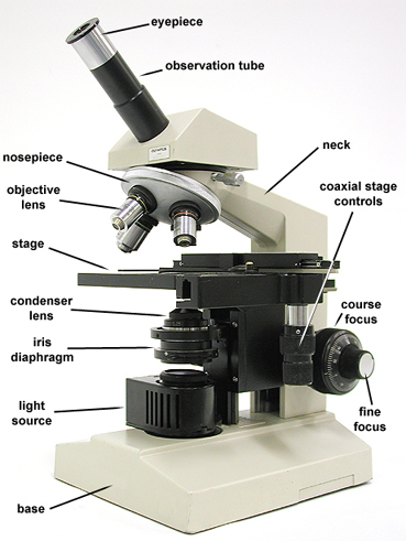
The lens closest to the observed object is called the objective. The lens that is on top of the tube is called an eyepiece. The magnification of the microscope is equal to the product (multiplication) of both lenses. For example, if the target has an increase power of 40 and the eyepiece has a magnification power of 10, then the object you observe will be magnified 400 times (40 times 10).
Composite optical microscopes are extremely useful for life scientists because they allow us to observe living microscopic organisms. This means that it is not necessary to kill an organism to observe it with this type of microscope. Other microscopes do not have that advantage.
When compound optical microscopes were invented, it was assumed that better lenses could be obtained with better lenses. But even the best compound microscopes can not increase in size more than 1000 times without erasing the image and losing details. Is it not possible to increase the objects more than 1000 times? Yes, but that increase does not depend on light.
Electron Microscopes
Certainly, optical microscopes are very useful, but their limited expansion power does not allow to observe very small objects; living beings as viruses or individual atoms and the molecules that make up matter. But these objects have been seen. How?
Much of the scientific research is done with the electron microscope. An electron microscope uses a beam of very small particles called electrons instead of light rays. (Electrons are some of the particles that make up an atom). The electron beam is not focused through a lens as happens with the optical microscope, but by magnets.
The objects observed with an electron microscope can be increased up to 1 million times. Through them, scientists can observe the smallest organisms, as well as individual atoms and molecules. Most of the time, the enlarged image is seen on a screen.
There are several types of electronic microscopes. You will read about two of the most common.
TRANSMISSION ELECTRON MICROSCOPE A type of electron microscope is called a transmission electron microscope (TEM). In a TEM, the electrons focus on an object like the rays of light in a compound microscope. The enlarged image, which can also be photographed, can be seen on a television screen. TEMs are useful for scientists to observe the interior of an object as structures within a cell.
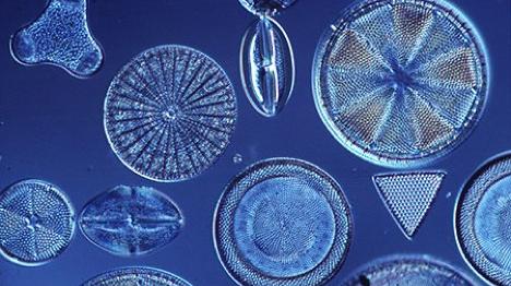
By using a TEM, scientists can study the internal structure of organisms such as diatoms. Source
SCANNING ELECTRON MICROSCOPE Another electron microscope is the scanning electron microscope (SEM). In an SEM the electrons focus on an object and are reflected (bounced) by it. The reflected electrons produce a three-dimensional photograph of the object. SEMs are useful for observing the external structure of an object, such as the arrangement of atoms in a solid.
The photographs taken by means of a scanning electron microscope provide surprising images. Source Pinterest
Both TEM and SEM present a problem. The object should be cut into very thin layers and placed under vacuum. This procedure implies that TEM and SEM can not be used to observe live.
Looking through the barriers
So far we have been looking for the use of microscopes to produce enlarged images of an object. Sometimes, however, scientists do not need to amplify an object, but look inside it. There are several instruments that allow "look through the barriers"
THE X-RAYS Do you know someone who has broken a bone? Maybe, yourself. A doctor could cut you to examine the bone, which would not be pleasant. There is a better way to do it.
For almost one hundred years, scientists have been using a type of radiation, X-rays, to see through objects. X-rays are similar to light rays, but we can not see them. They differ from light because they can easily pass through soft objects such as skin and muscles. But because they are blocked by dense objects such as bones, X-rays can be used to take pictures of bones inside an organism.
THE CT SCANS The computed tomography is a new invention that produces photographs of cross sections of an object. An X-ray machine inside a "CT scanner" takes 720 different exposures of an object. Each photograph shows a layer of the object. A computer analyzes and combines exposures in a photograph. Among its many uses, a "CT scanner" is used to photograph parts of the body, such as the brain.

Through a "CT scanner" it is not necessary for scientists to break bones to study them. Using a "CT scanner" they obtain a three-dimensional image of the inside of the skull. Source
MRI The magnetic resonance image (MRI) is another of the instruments that serve for inside objects. Produce images through magnetism and radio waves. Scientists can use MRI to study the structure of the cell body without damaging living tissues.

Magnetic resonance imaging helps scientists study the inside of the body. Source
In a future publication we will go further, and we will learn the most used instruments to explore the Universe. There are still many things to learn.
Previous articles
- Measurement and science - The metric system
- Discover the questions that scientists ask: Microscopic, macroscopic and global.
References website
https://es.wikipedia.org/wiki/Microscopio
https://es.wikipedia.org/wiki/Anton_van_Leeuwenhoek
https://es.wikipedia.org/wiki/Microscopio_%C3%B3ptico
https://www.news-medical.net/life-sciences/Types-of-Electron-Microscopes-Spanish.aspx
http://newsroom.gehealthcare.com/night-out-of-the-museum-mummies-in-the-ct-scanner/
I loved reading this. Microscopes comprise one of the best inventions of all time, considering its application. I will be waiting for the next addition to this series. Keep it up @osmerj
what science has increased is impressive. From the first microscope to the electron microscopy. In the last 50 years information has accumulated, which could be equivalent to the information that occurred in the previous 2000 years of history, without underestimating it. Recognizing the great advances achieved in previous years even without the current great technology
It is amazing the extent to which your post has shed more light on something that seem ordinary and somewhat trivial. For a non-science expert like me it's a learning point to read that a mere leather worker was the first to discover the existence of 'animalcules'
Your gyrating photograph of the MRI is frightening but educative by simply looking at it for a while. How far medical science has truly advanced!
Generally, I want to thank you for your inspiring post because it shows that anybody's name can enter the book of history in an area he knows next to nothing about:
Thanks @osmerj.
I love these comments, I'm glad you learned something new in my publication.
The image comes from a mummy from the National Museum of Archeology in Spain. It was found that one of them was pregnant, was between 20 and 35 years old and lived between 9 and 7 a.C. Science has really come a long way and there is still much to discover.
Thanks for your comment, friend.
I wonder how it feels when you are inside the MRI machine? :) really helpful post. looking forward to your next blog :)
I also ask myself the same question. Thank God I have never been inside a similar machine.
Really informative post
Buen trabajo @osmerj.
Gracias hermano
thank you, your post
I love science.
Such an amazing world living around us, so nice science allow us to learn more about it.
Great work!
Excelente contenido, la mayor de los exitos @osmerj