Nobel Prize for chemistry: Cryo-electron microscopy
Award for key method of Biochemistry
Insights into the molecules of life: Jacques Dubochet, Joachim Frank and Richard Henderson - three researchers who have developed cryo-electron microscopy - are awarded this year's Nobel Prize in Chemistry. This process has made it possible to see biomolecules in action and to represent them in atomic terms. We owe it to spectacular shots of bacteria in the attack on cells, photosynthesis molecules in the light, or the structure of the Zika virus.
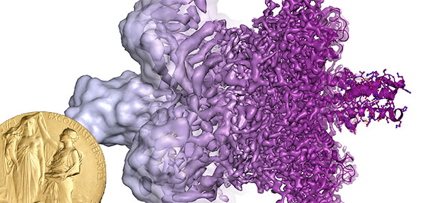
Cryo-electron microscopy makes unique insights into the structure of biomolecules possible. © Martin Högbom/ The Royal Swedish Academy of SciencesFor a long time, biomolecules such as proteins, DNA or RNA were the great unknowns of biochemistry. Although scientists were able to infer the structure of these molecules by means of X-ray crystallography, these images show the biomolecules only in crystallized, solidified form. How these complex connections move and how they interact with one another has remained largely unknown. Moreover, many biomolecules can not be crystallized and therefore completely withdrawn from our view.
Electron microscopy is also only suitable for the imaging of such biomolecules. For the rays to be reflected and thus to reveal its structure and shape, the sample has to be prepared, dried and, for example, steamed with heavy metal salts. However, this treatment alters many biomolecules and can even destroy them.
Richard Henderson: Sugar and low radiation
Richard Henderson was not satisfied with this situation. In his laboratory in Cambridge, he worked on a method by which he could visualize the photosynthesis protein Bacteriorhodopsin. His idea was to replace the water in the protein solution with a glucose solution that stabilized the molecule in the vacuum of the electron microscope.
To prevent the hard electron beams from destroying the sensitive protein, Henderson and his colleagues then tested how much information can be obtained with lower radiation doses. It was shown that by combining a lot of recordings, enough data can be collected about the molecular structure in order to obtain a first usable picture.
Based on this technique, Henderson and his colleagues first presented a 3D model of Bacteriorhodopsin in 1975, which showed how the protein chain of the molecule wallowed seven times through the adjacent cell membrane. Their images, with a resolution of 0.7 nanometers, were the best image of a protein ever produced by an electron microscope. But this was not yet a resolution down to the atom, and the sugar solution could not be used for all molecules.
Jacques Dubochet: cold shock for molecules
Jacques Dubochet found a solution to this problem at the European Molecular Biology Laboratory in Heidelberg. He discovered how molecules in aqueous solution can be kept in the vacuum of the electron microscope before being driven out. Even before him, researchers had tried to freeze the samples, but the ice crystals disturbed the electron beams and thus the image.
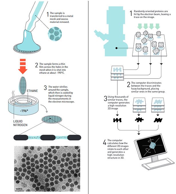
Schematic illustration of Dubochet's and Frank's method © The Royal Swedish Academy of SciencesDubochet's way was to cool the molecular solutions with liquid nitrogen so rapidly and radically that no ice crystals could form. Instead, the water solidifies to a glassy solid which does not break the radiation and thereby allows the image of the molecule enclosed in the vitrified water. In 1984, Dubochet and his colleagues succeeded in imaging viruses in solution for the first time - an important advance for medical research.
Joachim Frank: An algorithm makes the image sharp
The last step to the perfection of cryo-electron microscopy was made by Joachim Frank, who researched in the 1970s at the US Department of Health. He developed a software that can create high-resolution 3D images from several low-resolution electron microscope images. The computer does this by recognizing and combining recurring patterns in the molecular structure.
The decisive advantage is that the image information of several identical, randomly distributed and aligned molecules can be combined by this evaluation. As early as 1981, Frank was able to use these algorithms to generate first high-resolution images of proteins. In 1991, the researcher combined his software with the preparation method of Dubochet and was thus able to visualize the structure of a protein in 3D for the first time.
A new era of Biochemistry
In the meantime, by combining and improving all three methods, cryo-electron microscopy has greatly developed. Since 2013, proteins and other biomolecules can be precisely imaged down to the atom - in momentary snaps that freeze the molecules in the middle of motion or in action.
This method has brought biochemistry into a new era, says the Nobel Prize Committee. For a picture is often the key to understanding.
Source: Nobel Foundation, 04.10.2017 - DAL, The Royal Swedish Academy of Sciences

Congratulations! This post has been upvoted from the communal account, @minnowsupport, by n3bul4 from the Minnow Support Project. It's a witness project run by aggroed, ausbitbank, teamsteem, theprophet0, someguy123, neoxian, followbtcnews/crimsonclad, and netuoso. The goal is to help Steemit grow by supporting Minnows and creating a social network. Please find us in the Peace, Abundance, and Liberty Network (PALnet) Discord Channel. It's a completely public and open space to all members of the Steemit community who voluntarily choose to be there.
Sory friends help me.
https://steemit.com/aceh/@safrijals/rukok-tanyo-aceh-2017105t233144598z
This post has received a 0.35 % upvote from @drotto thanks to: @banjo.
**@n3bul4 got you a $3.33 @minnowbooster upgoat, nice!
Want a boost? Minnowbooster's got your back!
The @OriginalWorks bot has determined this post by @n3bul4 to be original material and upvoted it!
To call @OriginalWorks, simply reply to any post with @originalworks or !originalworks in your message!
To enter this post into the daily RESTEEM contest, upvote this comment! The user with the most upvotes on their @OriginalWorks comment will win!
For more information, Click Here! || Click here to participate in the @OriginalWorks writing contest!
Special thanks to @reggaemuffin for being a supporter! Vote him as a witness to help make Steemit a better place!
This post received a 1.6% upvote from @randowhale thanks to @n3bul4! To learn more, check out @randowhale 101 - Everything You Need to Know!
This post has received a 4.03 % upvote from @booster thanks to: @n3bul4.
Resteemed your article. This article was resteemed because you are part of the New Steemians project. You can learn more about it here: https://steemit.com/introduceyourself/@gaman/new-steemians-project-launch
Sory friends help me.
https://steemit.com/aceh/@safrijals/rukok-tanyo-aceh-2017105t233144598z
Amazing minds out there eh? I can't even comprehend half of it...lol