Basics of Reading an X-ray
G’day team,
I don’t think there’s a person alive who doesn’t think it’d be cool to be able to read an X-ray.
I remember being a kid and my sister coming home after breaking her arm, I remember bursting with excitement to see her X-Ray and anticipating an image of two bones jutting at obscure angles inside her skin. I was sorely disappointed. I saw nothing, more than that, I didn't even know what I was meant to be seeing!
And this is the frustration with X-rays for the average person, unless there’s a massive and clearly deformed bone, they’re not easy to understand.
So today I figured I’d go over some of the basics of X-ray, and start giving a bit of an introduction to what they are and how to read them. We’ll be mostly focusing on fractures because this is a good place to start with X-Rays and because unless you’re a Doctor this is probably all you’ll ever want.
If I have a lot of interest I promise to follow up with a more detailed one too!
So let's get going
A Tiny Bit of History
In December 1895 a Mechanical Engineer/ Physicist named Wilhelm Rontgen, experimenting with passing electromagnetic waves through different materials, decided ‘fuck it’ and threw his hand in front of the beam.
Bam!
The X-Ray was born!

Wilhelm Röntgen - Looking Dapper as Fuck
Image source
Later Wilhelm would take the first ever X-Ray picture, of his wife's hand, producing the famous image below!
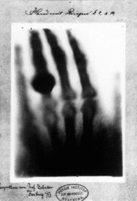
Image source
How's it Work
X-Rays work similarly to a regular camera. When you stand outside and a friend takes a photo of you, it’s because electrons are being shot at you by the sun, absorbed by your skin and clothing and then shot back out by your skin and clothing to eventually enter the camera where they record an image on a film.
With an X-Ray machine, the electrons are shot at an energy level far too high for the skin or soft organs to absorb, so they pass straight through. But bones are dense enough to soak up the electrons and when receptor board is placed behind that arm, we’ll get a picture of what the bones look like!
Kind of like finger puppets, but with bones!
Radiodensity
Awesome! One-hundred-and-thirty odd years have passed and thanks to generations of women and men much smarter than me, we’ve got highly specialized and incredibly accurate X-Ray machines. So what are they showing us?
Well the basics are the same. We shoot some X-Rays through the body and take a look at what’s going on inside.
What we see that is due to changes in ‘radiodensity’, or how many of those waves get through to the other side.
Things that come out looking white we call ‘radio-opaque’, these are structures that absorb radio-frequencies such as bone and metals.
Things that come out looking black we call ‘radio-translucent’, such as air and soft tissue (muscle, skin etc).
What’s Normal
Now, time to get practical.
The most important thing to understand, when looking at an X-Ray, is what normal looks like. Without a baseline, there’s no way anyone will be able to spot anything that might be wrong.
This means understanding basic bony anatomy and understanding what that anatomy will look like under X-Ray. It means understanding what ‘variations of normal’ may look like and what we expect to see in different age groups.
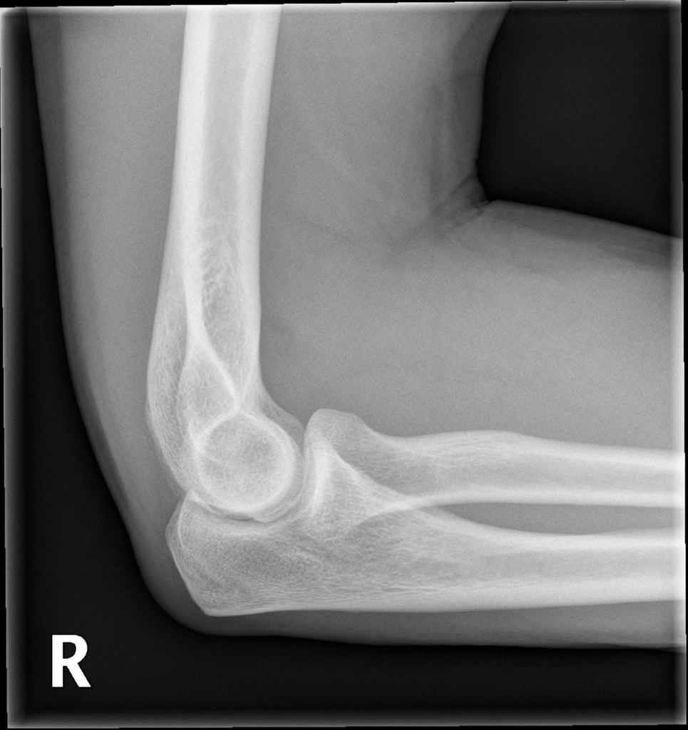
Let’s look at the X-Ray below…
So, let's go through some questions...
- What joint is this?
- Which side of the body?
- What angle are we looking from?
- How old is our patient?
- Is this image normal?
Well let's start at the top...
- This is an elbow
- This is the right elbow (this can be hard to tell on X-Ray, look for the tell-tale 'R')
- We're looking from a lateral view, that means from the side
- Here we really only need to break down between children, and not children. A child's bone will not fully 'ossify' (harden) till they finish puberty and till this age, the un-hardened bone will have see-through sections... so this is an adults bone.
- Yes it is normal, and let's look at why...
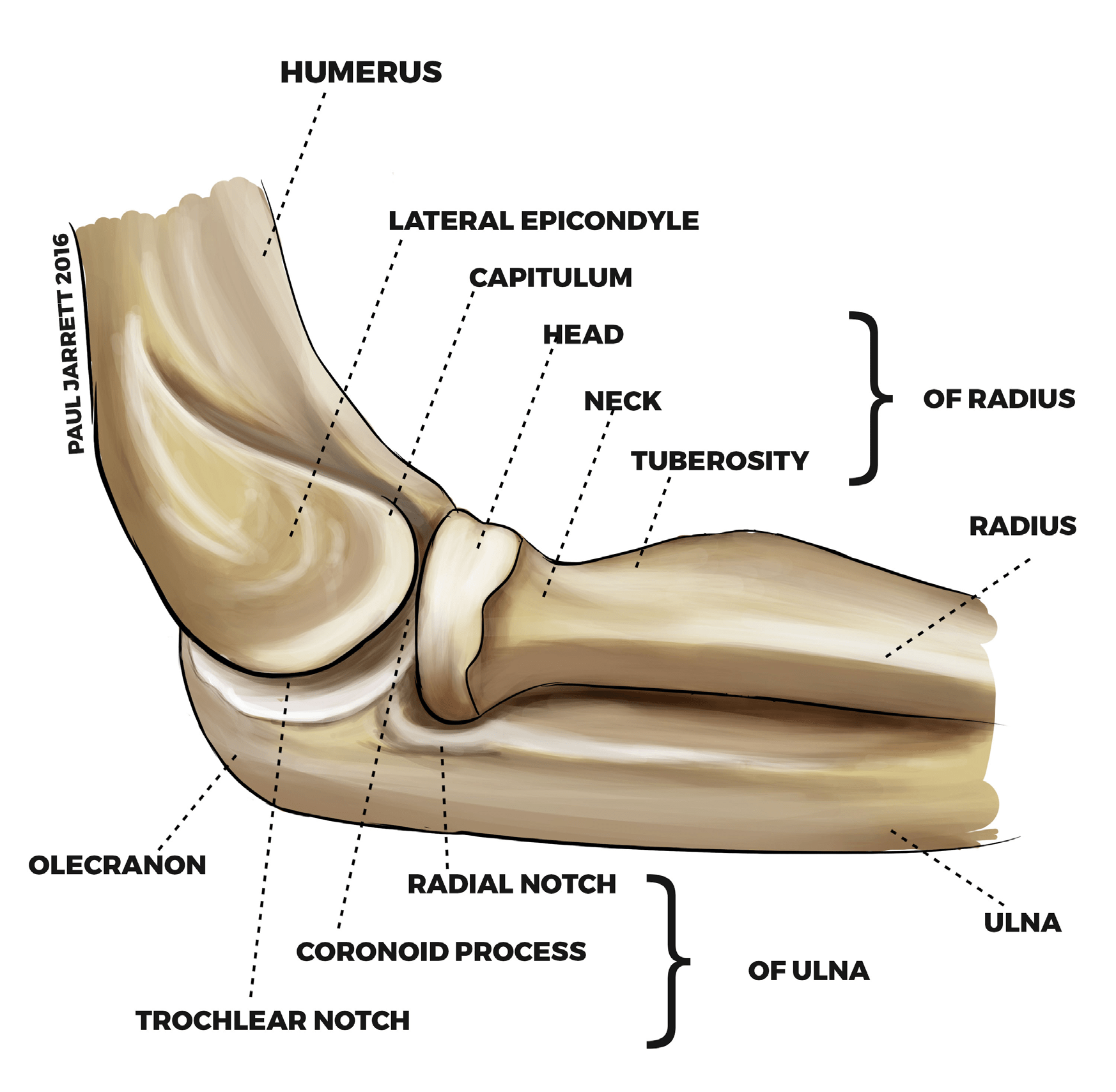
This is what we are looking at

Image source
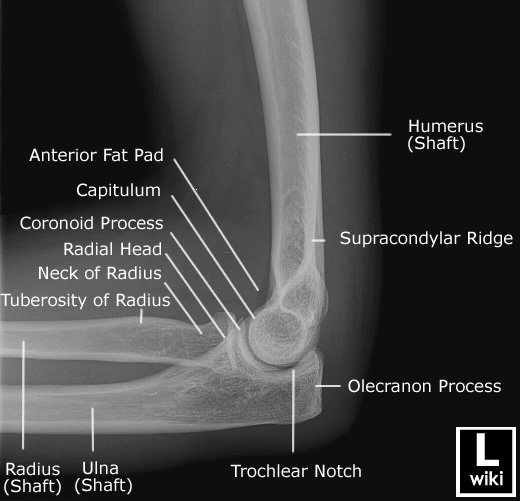
And what does this translate to? (sorry this one is flipped around)
As you can see, even understanding a simple elbow X-Ray requires a fairly in-depth understanding of bony anatomy.
Now we won't cover the anatomy of every bone and joint today, but if you're interested in peeking at a mate's, family members or your own X-Ray next time it might be a good idea to have a quick google of the region you're going to be looking at.
Fractures and Fracture Types
Okay so we're doing pretty well! We've got a basic understanding of what we're looking at, but now we need to know what we're looking for.
By definition, a 'fracture' (broken bone) is a disruption in the cortex of a bone. Now if we understand that a bone is made up of an inner spongy bone surrounded by a thick layer called the cortex (or compact bone), then a fracture is breaking of this cortex.
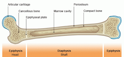
Image source
Let's discuss some types of fractures...
Comminuted Fracture
This is your classic 'well that's broken' fracture. It's defined as a fracture where the bone is in more than two fragments. Let's have a look below...
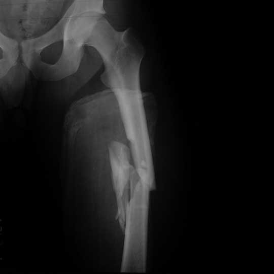
Comminuted fracture of the femur
[Image source](https://eorif.com/femoral-shaft-fracture-s72309a-
Nondisplaced fracture
It's as simple as it sounds! A fracture where the bone fragments are still in the right place, but they're... broken.
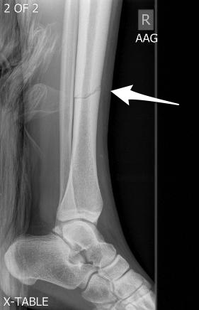
Nondisplaced fracture of the tibia
Image source
Spiral fractures
Time to get a little bit more complex. A spiral fracture is... nasty! Imaging your bone being held at both ends then twisted till it breaks... well that's a spiral fracture. The pattern we see in 3D is somewhat of a corkscrew style break, but in the 2D of an X-Ray this can be hard to pick up. What we see instead is a pattern that look s a little bit like a tan function...
Avulsion Fractures
Imagine you're five, your bones are soft because they haven't hardened yet, but you've got nice strong ligaments. You roll your ankle, badly, and something has got to give. In an adult it's usually the ligament, a sprain will swell up bad and hurt for a bit but the bone won't break. In children, because the bone is soft, the ligament often pulls part of the bone with it. We call this an avulsion fracture. The image below is a beautiful little example!
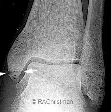
Image source
Buckle fractures
Another fracture often seen in kids. This time it's caused by pressure going down through a bone, causing it to 'buckle' in on itself, not unlike crushing a can. Here we're just looking for a bulging in the cortex of the bone. These 'bulges' can be tiny!
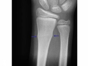
Image source
Bowing fracture
Again, generally seen in children, a bowing fracture is caused when a bone isn't so much brittle as malleable. As such when a force is applied the bone doesn't break like a stick of chalk but bends like a paperclip. Compare the top and bottom images below and note the clear bending of the bones in the lower image. For bonus points, can you spot the fracture in the top image?
Greenstick fracture
Kids again! Because they just have to make things so much more complicated! In a greenstick fracture there is disruption of the cortex, but not all the way through the bone.
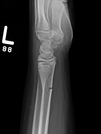
Image source
So there we have it, our types of fracture! Now who wants to put this all to practice?
Practice Time
Answers to come in comments section
Questions
Q1
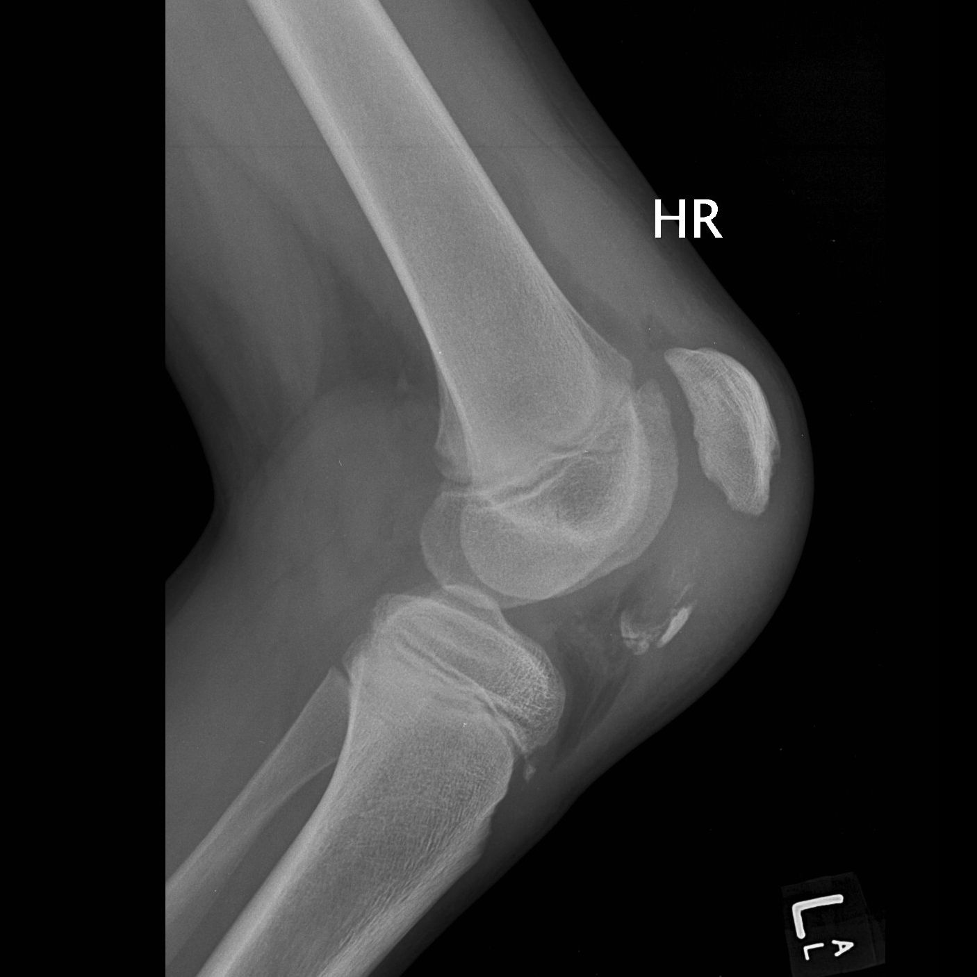
Can you find the fracture?
Q2
What type of fracture is this?
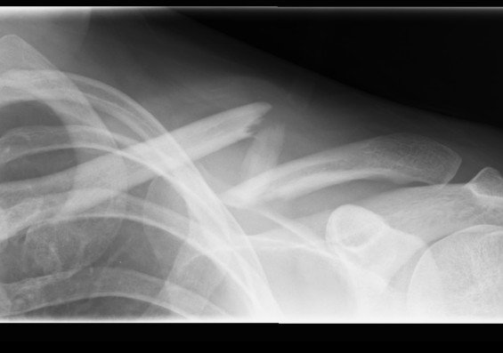
Image source
Q3
What sort of fracture is this?
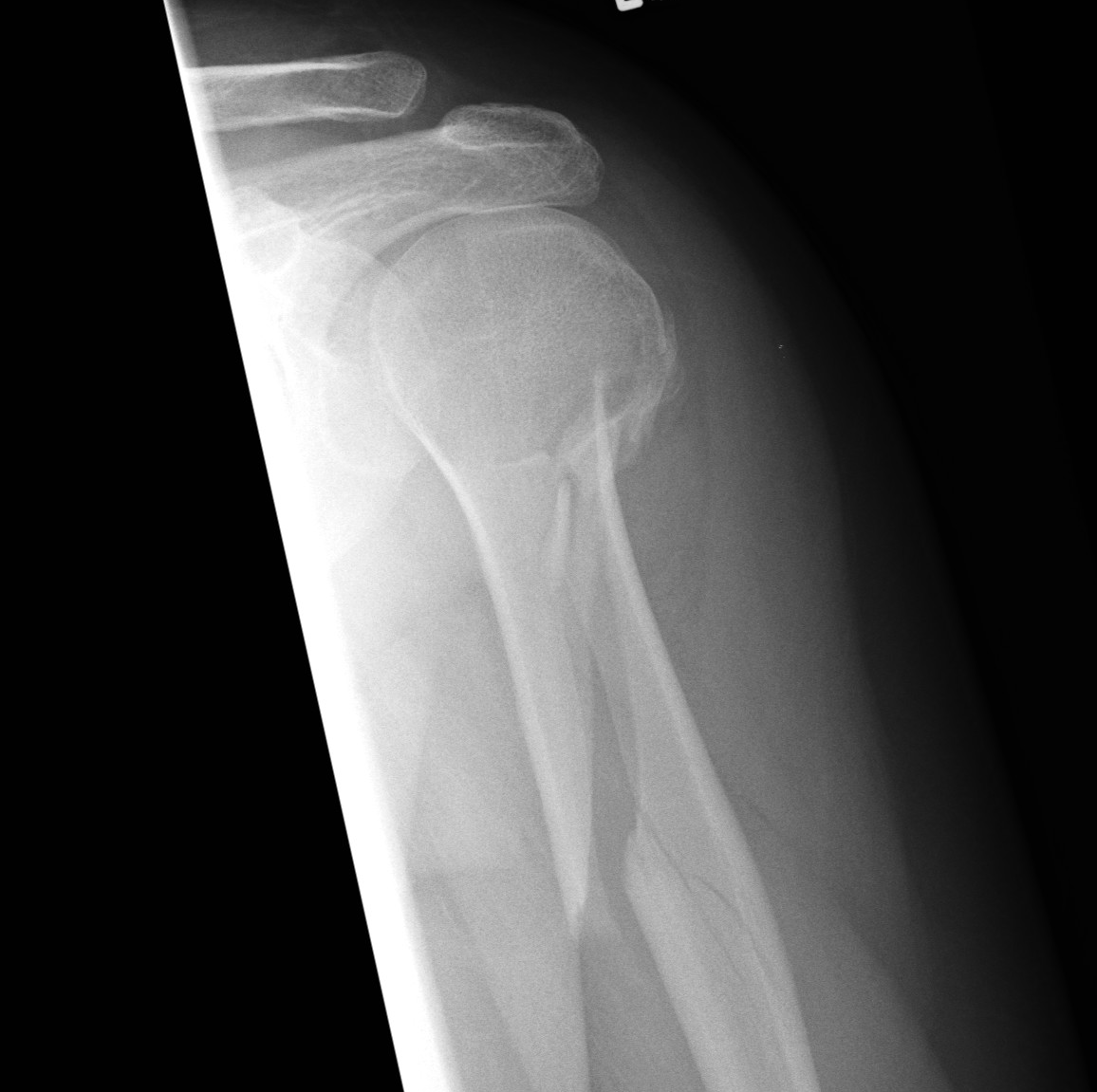
Image source
Q4
Where is the fracture here?
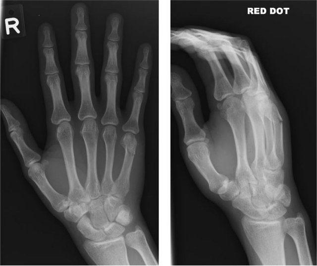
Image source
Q5
Bonus question (this may require some research) what sort of fracture is this?
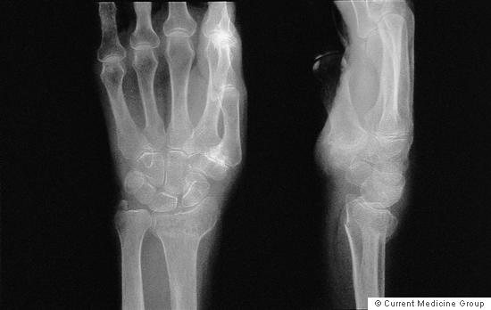
Image source
Answers will come out in the comments section in 24 hours!
That's it!
Thanks
So that's X-Rays
Hopefully, you've all learned a bit about reading X-Rays and now you can go out and impress everyone with your medical knowledge!
If anyone is interested I'm happy to keep posting questions regularly, just mention in the comments below!
Thanks again team!
-tfc
Resources
- Radiopaedia - great resource you can't go past to learn more
- Last's anatomy
- General Principles of Fracture Care
- OrthoInfo
- Innerbody
My Recent Posts
Hi, I'm Tom! (My life of medicine, science and fantasy)
Super Bugs - What is antibiotic resistance and how we are fighting it
Hydatidiform Mole – When Pregnancy Goes Wrong
Why is Antibiotic Resistance so Hard to Combat
Subconscious Discrimination - Test yourself using this Harvard University tool
Human Anatomy 101 - The Rotator Cuff


X-rays have become such a normality of our lives that I think everyone wonders about how to read them.
I'll do the easy question:
Q3 Spiral.
Great job! Here're the rest
An avulsion fracture of the patella (knee cap)
A comminuted fracture of the clavicle (collar bone)
Spiral fracture of the humerus
Greenstick/ non-displaced fracture of the 5th distal metatarsal
The metatarsal of th elittle finger, a little hard to tell which type of fracture it is at that resolution
A colles fracture of the head of the radius and ulna. Understand exactly what this is here
brrrr, I will dream of bones broken under an X-ray now...dammit.
Nice work!
Thanks mate... I find it's always nicer to see them under X-Ray than in person though :p
An avulsion fracture of the patella (knee cap)
A comminuted fracture of the clavicle (collar bone)
Spiral fracture of the humerus
Greenstick/ non-displaced fracture of the 5th distal metatarsal
The metatarsal of th elittle finger, a little hard to tell which type of fracture it is at that resolution
A colles fracture of the head of the radius and ulna. Understand exactly what this is here
good post friend greetings
Thanks mate
An avulsion fracture of the patella (knee cap)
A comminuted fracture of the clavicle (collar bone)
Spiral fracture of the humerus
Greenstick/ non-displaced fracture of the 5th distal metatarsal
The metatarsal of th elittle finger, a little hard to tell which type of fracture it is at that resolution
A colles fracture of the head of the radius and ulna. Understand exactly what this is here
Cool post, very informative and fun. Love the idea of quizzing at the end. Those were tough -- looking forward to the answers !
An avulsion fracture of the patella (knee cap)
A comminuted fracture of the clavicle (collar bone)
Spiral fracture of the humerus
Greenstick/ non-displaced fracture of the 5th distal metatarsal
The metatarsal of th elittle finger, a little hard to tell which type of fracture it is at that resolution
A colles fracture of the head of the radius and ulna. Understand exactly what this is here
Cool post, enjoyed this one.. :)
An avulsion fracture of the patella (knee cap)
A comminuted fracture of the clavicle (collar bone)
Spiral fracture of the humerus
Greenstick/ non-displaced fracture of the 5th distal metatarsal
The metatarsal of th elittle finger, a little hard to tell which type of fracture it is at that resolution
A colles fracture of the head of the radius and ulna. Understand exactly what this is here