What Biologists do all Day - Structural Biology AGAIN
In case you missed my last post about structural biology, click here to read it!
In short, structural biology tries to determine the structure of biological molecules (mostly proteins). That's more difficult than it might sound, as the parts of these molecules are too small to be seen with a microscope.
Last time, I showed you the last steps in determining a protein's structure, which wasn't so super smart, because now I have nice pictures for the first steps. But what can I do, these things happen.
After all, it's good for you, you get to look at very pretty protein crystals! Or at least I think they're pretty. I'm a huge fan of things like that.
A protein crystal that's used to determine the structure is made out of the same protein, over and over again, arranged in a symmetric structure. But proteins don't just do that by themselves. There are several methods to make them crystallize.
In this experiment, we used the "hanging drop" method, where the protein is mixed with some other chemicals (that control variables like pH and salt concentration) and then 1 µl (yes, microliter!) is put on a tiny class, flipped around and put on a well plate, which contains the same liquid the protein was mixed with. The protein we used is called "lysozyme", which is important for the immune system.@suesa
A well plate looks like this:
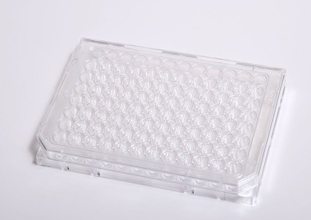
The holes are the wells. In theory, you can have a different protein-liquid mix in every well to test different conditions, because every protein has its own perfect conditions for crystal formation.
Over the span of a week, water evaporated from the tiny drop, which increased the protein concentration slowly and allowed crystals to form.
Under one condition, that happend fast, we could see so-called microcrystals after 30 minutes!
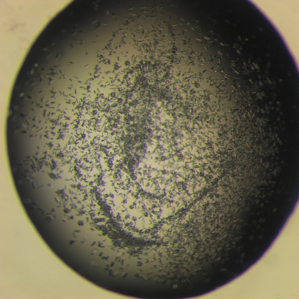
But these were totally useless. Equally useless were the needles that had formed in a different drop with different conditions after a week:
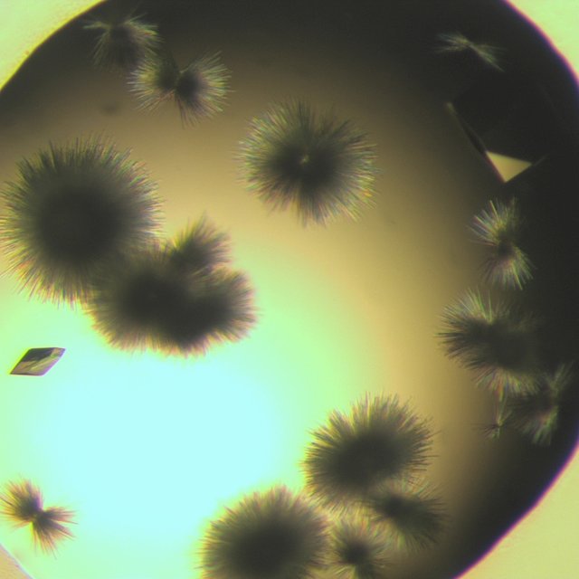
But to our delight, there were already proper crystals in this drop!
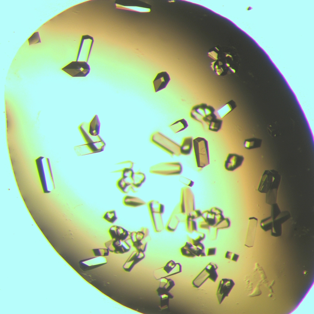
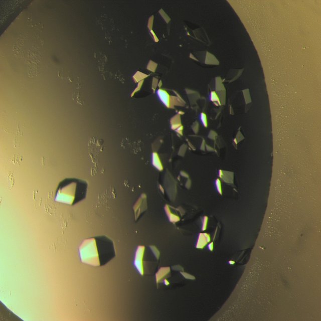
But they were too tiny for us to use. Other drops that looked great but were pretty useless:
%20B2%2050%20zp2.jpg)
%20d4%2050%20zp2.jpg)
%20c6%2050%20zp2.jpg)
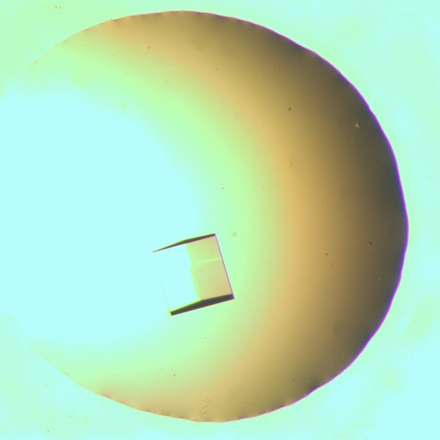
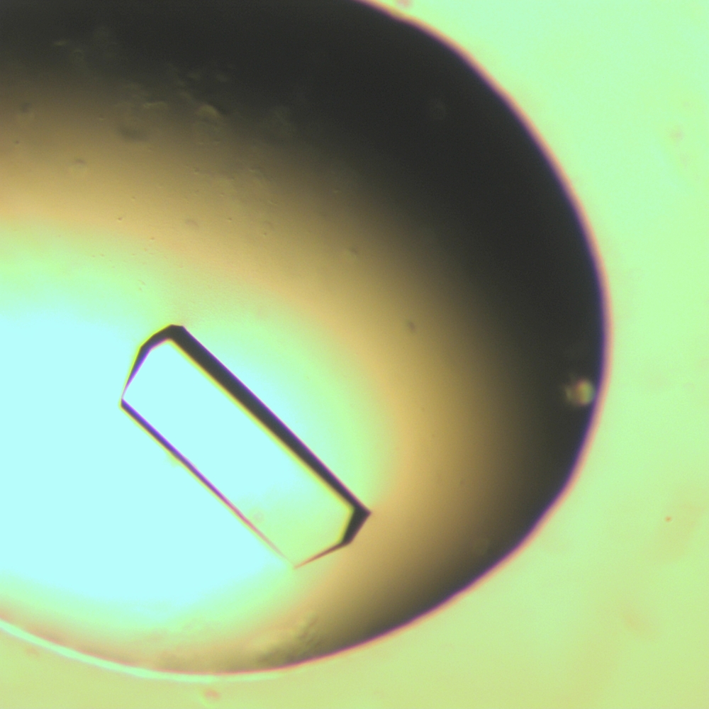
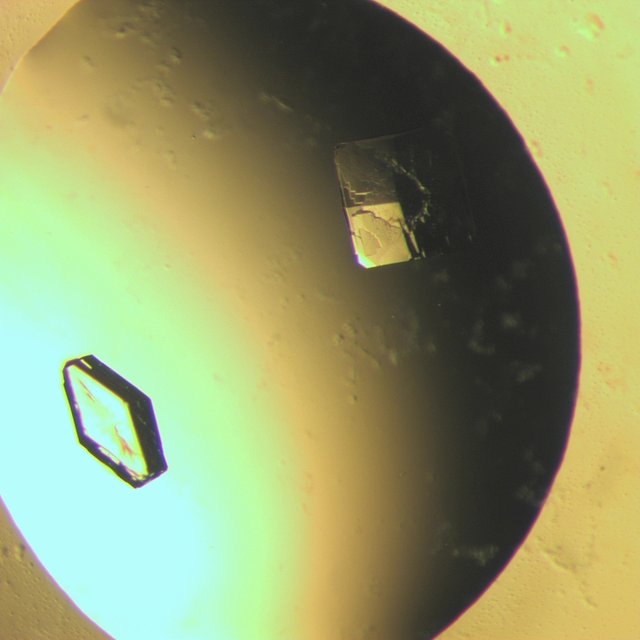
And finally, three drops that turned out great. If you look at them closely enough, you'll realize that they all have the same structure, we just captured them from different angles. That's because the protein was the same every time, which causes the same crystal structure, if there's a proper singular crystal.
Have you ever seen a salt crystal? Those look the same each time too, if you give them the correct conditions to develop!
%20d5%2050%20zp2.jpg)
The crystals, 400 µm to 900 µm big, were then fished out of the drop, using a tiny nylon loop and a microscope. Surgery is NOTHING against the precision needed here, because the crystals are very soft.
As soon as they're out of the drop, they're flash frozen in liquid nitrogen and kept at 100 degrees Kelvin. That serves to protect them, as they're put into this machine:
Where they're shot with x-rays. Those go through the crystal and are bent according to the protein's structure. A screen behind the crystal detects this, which creates the following pattern:
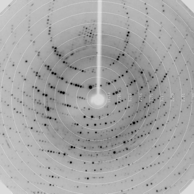
For this specific protein at least! If he had accidentally created a salt crystal (there's a lot of salt in the solution we used), the pattern would have looked more like this:
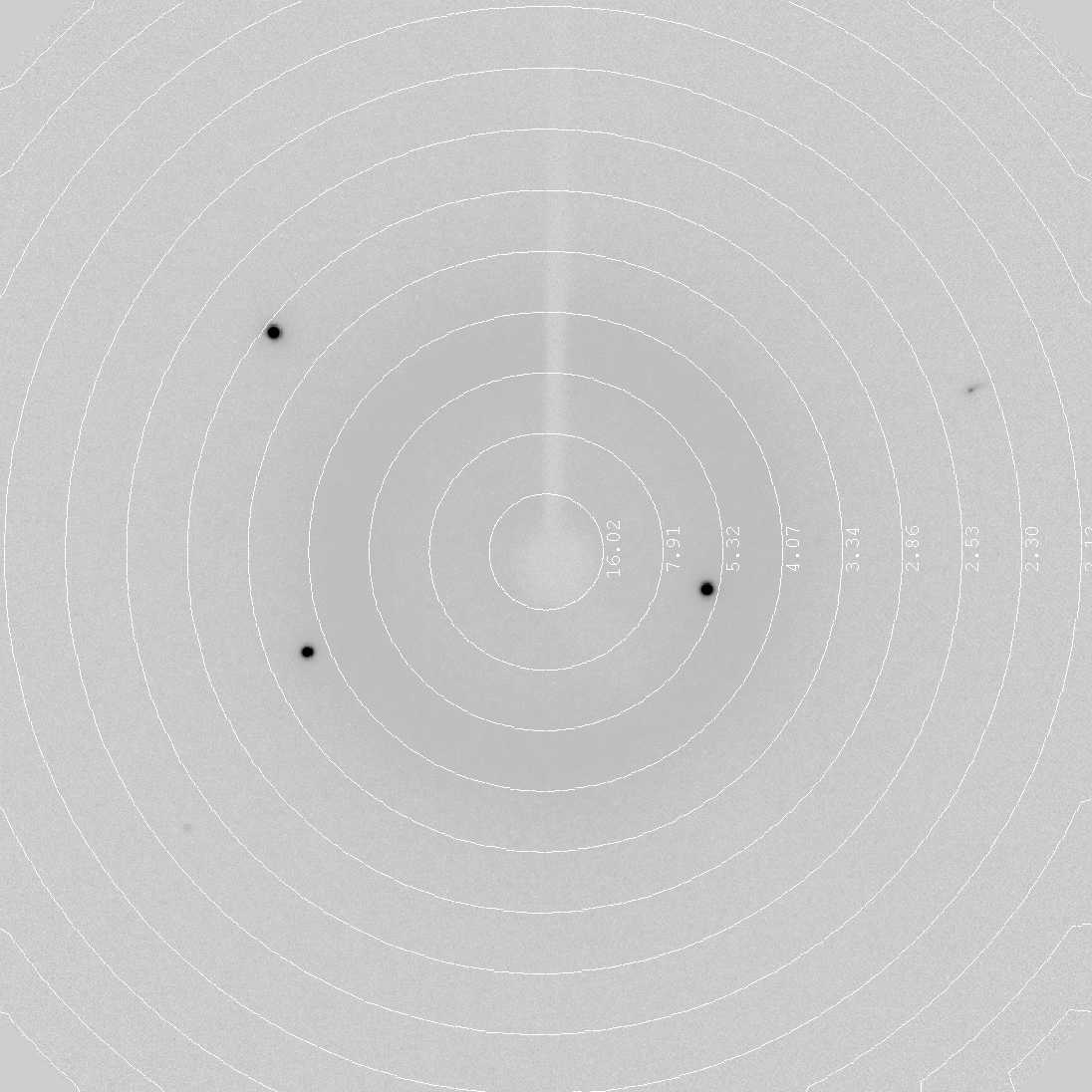
Why?
The more complex the molecule inside the crystal, the more complex is the pattern. The salt we used, NaCl (normal salt you eat) only has two different atoms. A protein consists of several amino acids which are all made up out of at least 4 different atoms (hydrogen, carbon, oxygen, nitrogen).
Not too long ago, a number of complicated calculations followed. Most of these can now be done by computers, which leaves us with the steps described in the post I published first and linked above. I really need to work on my timing!

I am licensing the pictures of the crystals under a Creative Commons 4.0 International License, as they're too pretty to not be shared!

Oh that's a nice post! The practical part of protein structure determination was completely missing during my studies.
I read your previous post and I am very impressed with your structure deductions from the X-ray diffraction. It really looks like black magic!
Will you reveal to us the magical conditions under which lysozyme formed the single useful crystal? :-)
There were several conditions. I'll be writing my lab report about it, can send you the table with the info if you want? Either steemit.chat or discord.
Sure, on steemit.chat :-) I'm just interested in what parameters matter for this kind of voodoo thing.
I remember the Biology classes although we only had analog microscopes.
Great job! The photos are amazing.
I am sorry I don't have more voting power.
A nice comment is often even better than an upvote :)
But I also voted :)
I appreciated the article!
Oh my, I know I've usually found very different feelings when reading your posts, but this time..
The engineer in me got a hard-on looking at this.
I don't care what it does, it's AWESOME.
I need one of those.
It shoots focused rays on a crystal while simultaneously cooling it to 100 K. The round screen catches the bent rays and transmits the pattern to the computer. XD
More Dark Magic i see...
Thanks for the post, i always wondered how they were able to do those 3d models of amino acids, now i know!!
It's dark magic in 60% of cases.
Been lovinggg the protein posts!!
Can I ask though, how do you get your pictures so clear?!
You must be taking the proteins and putting them back under a regular microscope right? I regularly struggle to take pictures of my microscope with my phone because I really don't know any better way to go about it then to hold up my phone and try to take a picture a shaky ass picture of a 0.4mm across lens. Honestly would kill to know how you get them so good
If your hands are shaking, try finding a way to attach the phone to microscope (like a phone holder which can be tightened on the microscope) and then have a timer or similar to snap the picture if possible, so touching the phone/camera will cause no shake.
The microscope has an integrated camera connected to a computer :P
Nooooooo I was hoping you wouldn't say that :( my uni needs new gear...
I've taken pictures like you described multiple times in the past, don't despair :P
Structural biology seems more fun than I’d imagine. Great shot of the protein crystals, microphotography is tricky work! I have a couple questions about those crystals. Are those “fuzz ball” crystals useless because you need a single large crystal to get a good defraction grating? Also are all the proteins the same composition,just with a different crystalline structure?
The "fuzz ball" crystals have no regular structure, which is absolutely necessary because only a symmetric crystal creates a pattern that can be used to calculate the actual structure.
I'm not sure what you mean by your last question.
We used the same protein (just different concentrations) and different pH/salt conditions. Some conditions produce nice, regular crystals, some don't. It depends on how fast the crystals grow (partly).
And structural biology is very, very tiring. It's not something I'd want to do all my life ^^
Hmm I think you answered my question with that last part. I’m not used to thinking about crystals made of proteins, so maybe my question about composition doesn’t even apply.
Here’s my thought process: minerals such as pyrite and marcasite are polymorphs of one another. This means they have the same composition (Fe-sulfide), but different crystalline structures. The difference in structure, caused by differences in growth conditions, distinguish the two as unique minerals.
When I saw your crystals, I thought, “maybe those proteins are polymorphs?” I’m not sure if polymorphs is even the right word to use tho 😅.
Thanks for the reply, interesting stuff!
Think I don't know enough about rocks to make give a good answer to this :'D
Growing cystals is kind of a dark art scientifically speaking. Results are unpredictable and seldomly repeatable.
One rare occasion where doing the same thing and expecting a different outcome is not madness :D
I joked that we're doing dark magic while we were looking at the crystals and comparing conditions. One of the supervisors looked me dead in the eye and was like "Don't tell anyone, they'll just start burning us again".
Very concise and thoughtful article!
I have one question tho, as a result protein-liquid mix you either get protein or crystal - or is it crystal every time (given enough amount of time).
And all the crystals look like building blocks of replicators from Stargate SG-1 :)))
Cheers!
P.S.
When I was in high school it was a tough call between biology and astrophysics. I went for astrophysics, but still love biology before all other sciences.
There are several things that can happen. The protein can just fall out and not crystallize at all. Different forms of useless crystals can form (like the needles). Or ... no crystal at all:
Thanks for clarification. :)
Many proteins are impossible to crystallize at date, i´ve tried 4000 mixes on a single protein with out a trace of crystal :)) 4000 is not so much thou its just 40 x micro arrays with 100 different crystallization spots/mixes(as seen in the article) and a robot does all the work so just pray for crystals :)
What instruments do you use to measure the sizes of the crystals?
We have a microscope that's connected to a computer, which has a picture recording and a measurement program. If we feed the measurement program with the picture and the magnification we used, we can just draw a line from one end of the crystal to another and it gives us the size.
I hate biology lessons when I was in junior and senior high school because my teacher just told me to record and memorize the names of Latin hard to remember.Now I realize there are many interesting and useful things in the science of biology. if the teacher taught me in an easy and convenient way, maybe I would be interested in becoming a doctor:)