How Do Skeletal Muscles Contract??
Want to know a fun fact? You've been lied to your entire life.
That's right. Everything you know is built upon a facade.
What is it this time? Aliens? Talking dolphins? Did Stella really not get her groove back?
Nope. Not even close...
Ready? Prepare yourself...
Muscles. Don't. Flex.
Okay, so maybe it's not the biggest lie ever told, but you gotta admit that you've probably thought it was true your entire life.
I guess the only question now is, what exactly is it that muscles do?
The answer is very simple — they contract. But what exactly is a contraction?
To figure this out, we need to learn some of the basic anatomy of muscle tissue.
3 Types of Muscle Tissue
If you recall from a previous post about muscle tissue, there are 3 different types of muscle in the body:
Smooth and cardiac muscle are both involuntary, meaning that you can’t decide when they contract. Smooth muscle is found all throughout the body, including inside of blood vessels and your digestive tract.
Cardiac muscle is found exclusively in the heart, and is some of the most unique tissue in the body.
Skeletal muscle however is voluntary, meaning that you can decide when to contract it. This is the type of muscle most of us think of when we hear the word “muscle".
To make things easier, we will only be speaking about skeletal muscle from here on out.
At a later date, I will delve much deeper into muscle tissue in general, but specifically smooth and cardiac. For now, simply understand that they use similar, yet different processes in their contraction.
Either way, they contract.
Skeletal Muscle
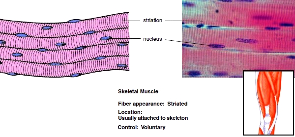
Skeletal muscle is what we call multi-nucleated, meaning that one individual cell has many nuclei that assist in its cellular functioning. Skeletal muscle cells can also be very long, bordering on multiple feet in length at times. A good example of this would be the Sartorius muscle, which depending on your height, can obviously reach differing lengths.

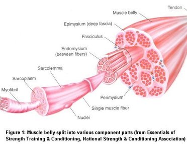
Let's Look Inside
If we zoom into an individual skeletal muscle cell, we find even smaller structures called myofibrils. Myofibrils are made up of the functional units of a muscle cell, called sarcomeric units, or sarcomeres, which are stacked end-to-end throughout the length of the myofibril.
Sarcomeres are defined as the region between the Z-Lines, Z-Membranes, or Z-Discs. Each name corresponds to the same structure, so whichever one you prefer to use is acceptable. The Z stands for the German word Zwischen, which literally means between.
As with every other biological system, if work is going to be done, proteins are required. The proteins of muscle tissue are myosin and actin.

Myosin consists of a head and a tail/rod. When several myosin proteins are stacked onto one another, the structure is now called the thick filament.
Actin molecules are tiny little bead like proteins, that when stacked together form a long and slender structure called the thin filament.
Myosin and actin are so extremely attracted to one another, a protein complex is required to block the receptor/attachment sites on the actin molecules, otherwise the muscle would be in a state of permanent contraction.
This protein complex consists of two proteins:
- Troponin
- Tropomyosin
Tropomyosin is another slender protein that acts to cover the receptor/attachment sites. To do this, troponin binds tropomyosin down to the thin filament.
Time to Contract
In order for a muscle contraction to occur, calcium ions are diffused into the skeletal muscle cell. For as much as troponin loves tropomyosin, it enjoys the presence of calcium infinitely more so.
Calcium will then bind to troponin, causing a change in conformation, and thus relaxing its grip on the tropomyosin. As the tropomyosin starts to relax, the binding sites on actin become available, and myosin takes advantage.

The myosin heads will now bind to actin, and a change in conformation will occur. This results in what’s called the power stroke.

It’s these power strokes that bring the Z-lines closer together, or what we can now call a contraction. When you add up the hundreds of thousands of power strokes, the end result can be quite dramatic.

All Good Things Must Come to an End
In order for a muscle contraction to stop, a few things need to occur. First, ATP (adenosine triphosphate) needs to bind to the myosin head and hydrolyze, becoming ADP (adenosine diphosphate) and a spare phosphate. When this occurs, the myosin head is “cocked” back into a position of high energy, ready for another power stroke.
Next, the calcium ions need to be removed from the troponin. This is accomplished by ATP binding to the calcium, and hydrolyzing into ADP, effectively "pushing" the calcium out of the cell. When this occurs, troponin once again binds to tropomyosin, blocking the receptor sites on the actin molecules, and the muscle is now in a relaxed state.
There's Plenty More to This Story
As they say, this is only the tip of the iceberg. In upcoming posts, I'll be diving much deeper into all 3 muscle tissue types, the different types of contractions, muscle cramps, and even how the brain sends the signal to contract in the first place.
So then, what is a contraction?
Contractions are a muscle generating tension. When calcium ions are introduced into the skeletal muscle cell, they bind with the protein troponin, which is "holding" the protein tropomyosin down against the thin filament, blocking its receptor sites. When the receptor sites become available, myosin will bind to the actin, and a power stroke will occur.
The fact is, flexion is what we call a joint action. Muscles can cause flexion to occur, but they don't flex themselves.
For example, when you are doing the typical biceps curl, your elbow joint is flexing. Now, the contraction of biceps brachii caused the elbow to flex, but the joint is the only thing flexing. Not the muscle.

It may seem like we're being nit picky, but if we want to truly understand the body, we need to be.
So next time someone says to "flex your muscles", feel free to shatter the facade!
See ya next time!
If You Enjoyed This, Here Are a Few More You Might Like
Let's Learn Anatomy #1
Let's Learn Anatomy #2
Let's Learn Anatomy #3
Let's Learn Anatomy #4
What Is a Heart Attack?
Why Are Human Babies So Incredibly Weak???
Why Is Your Butt So Big???
Evolution, Society, and Cryptocurrency... How Does It All Relate???
What Is Asthma?
What's the Difference Between a Strain and a Sprain?
Who knows where the red pill will take us...
thanks for writing a great post :) These are helpful for me preparing for the MBLEX .. It threw me at first because the image of the Quads was missing the Rectus Femoris.. but that gave me hope for the test since I noticed it was missing.. lol .. I have been a CMT for years.. relocated to new State
Thanks! I was thrown off by the picture myself. What's funny is that it's definitely missing Rectus Femoris, but it still looks like it. Vastus Intermedius has a different fiber orientation than what is presented. Oh well! All we needed was the Sartorius!
I'm glad to hear you're feeling ready for your licensing exam. Good luck!
Still a few months out I have to apply give them some money I went to school so many years ago fortunately I worked in medicine so some of it I remember from that by the way nice to meet you
It's very nice to meet you as well!
Nicely explained! How protein move along actin is really amazing.
I was told that dyneine was the motor protein which move along microtubules in cells. Their function looks quite similar haha~
Yes! Dyneine deserves a post in its own right. The way that cells move proteins and facilitate their own movement through their environment is astounding.
Absolutely brilliant stuff man! This is an excellent explanation on muscular form and function, even focusing on some of the more indepth (and important) concepts such as ION Transport across the Sarcolemma. This would make for an excellent summary of muscular function for a First Year Health Science course or Anatomy course.
Thanks! Muscle physiology is endlessly fascinating. It was an immense struggle to write this simply because I wanted to include much more than I did. I'm going to need to write another post that includes the neuromuscular junction and corticospinal tract.
It really is and there is a shit tonne of information regarding neural signal transport and muscular response, the NMJ and Corticospinal structures are incredibly important, I also find that discussing how nerves are descending (Which I mean that the nerves along the spine that correspond to a particular muscle group do not align perpendicular so that the Nerve origin is always more superior relative to the muscular group's nerve ending), and hence why Spinal Taps are typically done in L4-L5 to avoid paralysis/nerve damage of organs and any higher structures. Also I think the Cauda Equina is a pretty awesome point to show explicitly the density of nerves packed into the spinal cord.
I couldn't agree more! You have me sold! I'll be writing a post on the pathway motor neurons take as they descend from the motor cortex to the terminal end of the axon here soon!
I appreciate your enthusiasm and input! Thank-you!
Congratulations @justincottle! You have completed some achievement on Steemit and have been rewarded with new badge(s) :
Click on any badge to view your own Board of Honor on SteemitBoard.
For more information about SteemitBoard, click here
If you no longer want to receive notifications, reply to this comment with the word
STOP