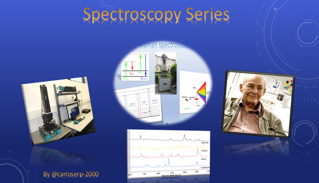
Spectroscopy Series VOL. 6: Raman confocal microscopy
Happy new year scientific community of steemit and especially steemSTEM today I will share the 6th part of my spectroscopy series.
If you want to read about the previous deliveries click on the following links:

One of the techniques of the Raman Spectroscopy that is perfectly adapted to this, is the confocal microscopy, since it allows to increase the contrast, definition of images and in turn build and obtain three-dimensional information.
Through this equipment we can observe spectra of chemical and biological samples. The invention of the confocal microscope in principle was for the study of biological and neurological events that occur in vivo, this was thanks to the scientist Marvin Minsky whose main objective in his study was to obtain information from neural networks using uncolored preparations of living brain tissue.

Marvin Minsky Licensed CC BY 3.0
The main objective of the patent created by Marvin Minsky was to improve the functions he had at that time in a fluorescence microscope, that is, to create a wider image to be able to observe at any nanometric scale any type of object, mainly very small organisms such as cells. The microscope consisted basically in irradiating the sample uniformly with a light source and thus occupying all the capacity of the material without wasting any portion of the sample.
The difference between the fluorescence microscope and the confocal microscope is that the optical path of the light was excited at the same time and the fluorescence was detected by the photodetector in the microscope chamber, whereas the confocal uses the light and a small hole plane conjugate optically with the detector and thus be able to emit the signal that is outside the focus of the lens and hence the name "confocal".
The advantage of creating this type of technique is in principle to have an exact focus with a confocal image using an aperture or hole the size of a pin, allowing a very sharp definition of the image of the thickest samples of the focal plane of the microscope.
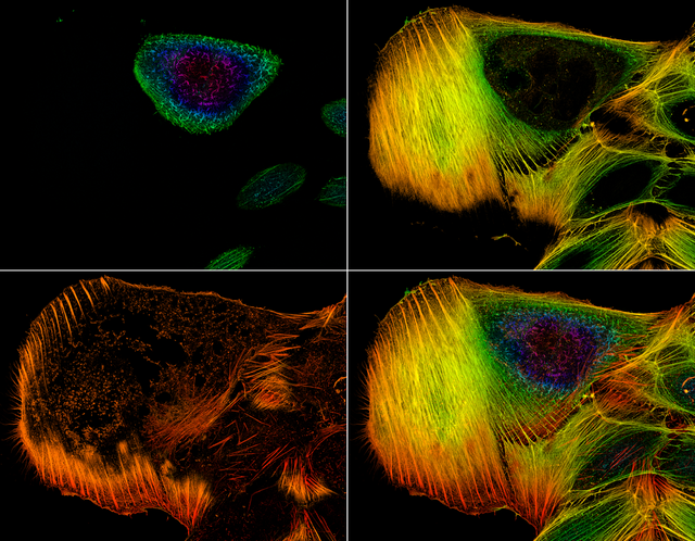
These are some examples that show the distribution of actin filaments in a cell through the confocal microscope Licensed CC BY-SA 4.0
With the passage of years and different technological advances this microscope was becoming very popular, being used by different scientists from different areas to study live samples and obtain three-dimensional information from them. Today we use it for chemical analysis of material samples and it is perfectly suited to spectroscopy due to its simple configuration.
We know that Raman spectroscopy is very well coupled to the confocal microscope, allowing us to obtain a spectral spectrum of some specific specimen in each pixel of the image. This type of imaging system has the ability to have a nanometric resolution of any material.
We can obtain spectral information using different spectroscopic techniques such as, for example: absorption, transmission, reflection, photoluminescence, emission, fluorescence and of course Raman. Undoubtedly, the Raman technique is preferred by many scientists because it is based on the inelastic dispersion of monochromatic light when in photons its frequency changes at the moment of interaction with the material.
The photons of the laser light are absorbed by the sample and re-emitted later. The frequency of the emitted photons is shifted up or down compared to the original monochromatic frequency, which is known as the Raman effect. The Raman change provides information about the vibrational and rotational energies of the molecular bonds.
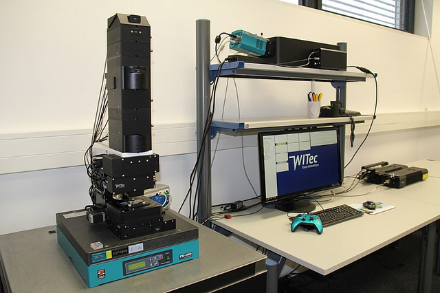
Raman confocal microscope Licensed CC BY-SA 4.0
Currently, there is a great diversity of advanced confocal microscopes that can be coupled to spectroscopy, allowing users to study samples with rough and irregular surfaces, since during the scanning of the sample an optimal focus can be obtained in real time through a video with White light. All this allowing greater ease of handling to the users, since manual focus is not needed.
For several years it could be observed that the confocal Raman spectroscopy technique was the indicated one to determine the energy levels and vibrational modes within a molecule, this has the great advantage that with only excite a small part of the molecule changing the length of excitation wave, we could have this kind of information very accurate. The simple fact of adapting a Raman spectrometer to a microscope gives us a very controlled sample volume, by which I mean that we only need a spatial filter to perform the analysis of the sample.
Nowadays different methods are used for some more practical, others not. However, Raman spectroscopy adapted to the confocal microscope gives us the possibility to analyze individual particles with dimensions less than 1μm and this is fabulous because no other method has this capacity. In addition to mentioning all this we can not forget that for this technique you do not need any type of preparation for the samples that you want to analyze, this allows the user a time saving and of course reusing the sample for later analysis because it shows them to your they are not destroyed.
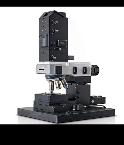
Confocal Raman microscope alpha300 R for Raman images and 3D volume scanning Licensed CC BY-SA 4.0
Important to emphasize about Confocal Raman spectroscopy is that it shows us spectra with sharp bands and very characteristic in the specific molecular bonds of each sample of the material analyzed, that is, it is being like its fingerprint.
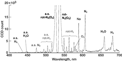
Raman spectrum in wavelength scale Licensed CC BY-SA 3.0
The intensity of the bands in a Raman spectrum is proportional to the concentration of the corresponding molecules and, therefore, can be used for quantitative analysis.
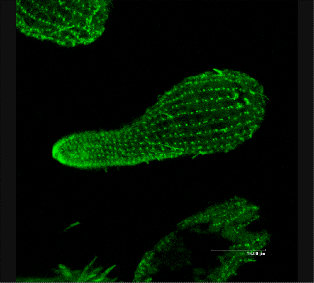
Beta-Tubulin in Tetrahymena cell, visualised using GFP-marked anti-beta tubulin antibodies under confocal microscopy Licensed CC BY-SA 3.0
Although in the analysis of certain types of samples, such as cells and tissues, the spectral characteristics give us information about certain cellular components, it allows us to specify in detail the qualitative analysis and discriminate between very similar materials.
Some studies that have been done with this technique are in biological samples as mentioned in the first part of the post, specifically in the identification of cells, allowing the bands to stand out and thus be able to identify regions of the cell membranes.
Applied studies in materials sciences such as graphene. In the same way applications in the pharmaceutical industry in the manufacture of different types of medicines.
Conclusion
In summary this technique offers many advantages since the preparation of the sample is extremely simple, it does not require a specific preparation, it contains a non-destructive test, we can identify phases directly, in turn we can perform analyzes to crystalline and amorphous solids and of course biological materials, allowing to identify the properties of each material in a three-dimensional image with a high resolution, in addition the range of variation of the temperature is between 250 and 600 ºC.
If you want more information about the subject you can visit the following links:
Difference between confocal and fluorescence microscope?
A practical guide for fluorescent confocal microscopy
What is the difference between PL(Photoluminescence ) spectroscopy and Raman spectroscopy ?


Video credits @gtg
This post has been voted on by the SteemSTEM curation team and voting trail in collaboration with @utopian-io.
If you appreciate the work we are doing then consider voting both projects for witness by selecting stem.witness and utopian-io!
For additional information please join us on the SteemSTEM discord and to get to know the rest of the community!
Hi @carloserp-2000!
Your post was upvoted by Utopian.io in cooperation with @steemstem - supporting knowledge, innovation and technological advancement on the Steem Blockchain.
Contribute to Open Source with utopian.io
Learn how to contribute on our website and join the new open source economy.
Want to chat? Join the Utopian Community on Discord https://discord.gg/h52nFrV
I love this, and I need to go back and read the the previous volumes. Raman is perhaps my favorite form of Spectroscopy. I have no experience with it, but I do with IR, FTIR, UV-VIS, FLourescence and NMR. I think what I like the most about Raman, is its more simple design versus its ability. From the perspective of service engineer and soon instrument designer, simple is good. The chemist in me likes the ease of sample prep. Thank you for sharing, and you may want to check out my blog on TEM. Resteeming.
Greetings soon I will be publishing about FT-IR, ultraviolet-visible and other technologies that are based on the Raman spectroscopy.
Without a doubt for physicists, chemists and engineers specialized in material science, Raman is extremely important.
I'll go through your blog, thanks for your comment @roguescientist84