A brief history of ultrasound elastography - Part 1
This is the first part of a very brief history of ultrasound elastography. I would like to start with a short general introduction and a brief discussion of Vibration elastography imaging. If you have any questions, please let me know in the comments below.
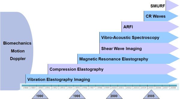
As early as 1952 research has been conducted in the area of shear waves, longitudinal waves and surface waves propagation in soft tissue [1]. However, the findings of this research did not get much feedback from the scientific community at that time. The fact that mechanical waves in soft tissues can propagate with speeds 100 times slower than the speed of sound in air appeared to be hard to accept [2]. For almost three decades these theoretical findings were not followed by any practical applications until two first research papers, looking into the possibility of ultrasound elastography measurements, were published almost simultaneously in 1982 [3] [4]. In 1983, a paper by Fujimoto et al. was published. That paper seems to have been the first to describe the use of dynamic tests to ultrasonically estimate the compressibility and mobility of breast tumours [5]. Due to the fact that this paper has only been published in Japanese, its impact on the English speaking research community has been limited and it was not until 1988 before follow-up research papers based on the ideas presented by Fujimoto et al. were published. Since 1988 many technologies for elastographic imaging were developed. An overview of some selected developments is given in the figure below.

Elastographic imaging developments from 1988 to 2008 (Source: [6])
Vibration elastography imaging (1987)
Vibration Elastography Imaging or Vibration Amplitude Sonoelastography is an ultrasound technique in which an external vibration in the low frequency range of 20 to 1000 Hz is applied in order to excite internal vibrations within the examined tissue [6]. Detection algorithms known from conventional Doppler mode ultrasonography can be employed to acquire a real-time image. This method was invented in 1987 [7] and was subsequently further developed. Further research discovered that, in some organs, additional information (e.g. shear wave speed) can be deduced from modal patterns created by external vibrations [8]. Implementing various time and frequency estimators make this a real-time diagnostic method with applications to superficial and deep organs [6].
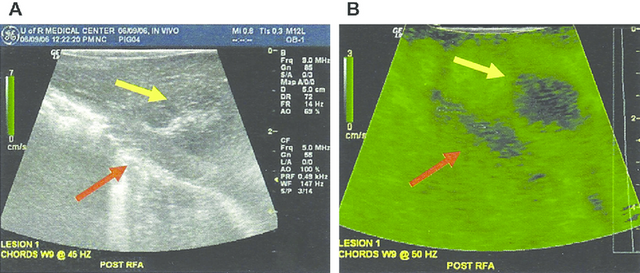
The figure below shows a sample application in which the liver of a pig with a thermal lesion has been examined in vivo.

Lesions (yellow arrow) of a pig liver in a normal B-mode scan (a) and as sonoelastography image (b). The red arrows indicate the boundary of the liver. (Source: [6])
One advantage of Vibration Elastography Imaging is its compatibility with most color Doppler scanners. However, this technique produces only relative image contrast related to local hardness. Thus, a uniform background within an organ is needed which can be difficult to obtain. Additionally, the ultrasound system also has to be equipped with a vibration source which increases manufacturing costs and the size of the system [6].
References
- Henning E. von Gierke et al. “Physics of Vibrations in Living Tissues”. In: Journal of Applied Physiology 4.12 (1952), pp. 886–900. ISSN : 8750-7587.
- Armen Sarvazyan et al. "An overview of elastography - an emerging branch of medical imaging". In: Current medical imaging reviews 7.4 (2011), p. 255.
- L.S. Wilson and D.E. Robinson. “Ultrasonic measurement of small displacements and deformations of tissue”. In: Ultrasonic Imaging 4.1 (1982), pp. 71 –82. ISSN : 0161-7346.
- R.J. Dickinson and C.R. Hill. “Measurement of soft tissue motion using correlation between A-scans”. In: Ultrasound in Medicine & Biology 8.3 (1982), pp. 263 –271. ISSN : 0301-5629.
- PN Wells and HD Liang. "Medical ultrasound: imaging of soft tissue strain and elasticity". In: Journal of the Royal Society (Nov. 2011).
- K J Parker, M M Doyley, and D J Rubens. “Imaging the elastic properties of tissue: the 20 year perspective”. In: Physics in Medicine and Biology 56.1 (2011), R1.
- RobertM. Lerner et al. “Sono-Elasticity: Medical Elasticity Images Derived from Ultrasound Signals in Mechanically Vibrated Targets”. English. In: Acoustical Imaging.
Ed. by LawrenceW. Kessler. Vol. 16. Acoustical Imaging. Springer US, 1988, pp. 317–327. ISBN : 978-1-4612-8051-4. - K. Parker and R. M. Lerner. “Sonoelasticity of organs: shear waves ring a bell”. In: Journal of Ultrasound in Medicine 11.8 (Dec. 1992), pp. 387–392.
Thanks a lot for your attention and stay tuned for the second part of this series. As a reward for reading this article and in anticipation of learning more about the SMURF technology, I present to you:

Papa Smurf. Source: https://en.wikipedia.org/
I never realized there were so many different ultrasonography techniques. I sort of only thought there was one... Will look forward to the next installment in this series.
Add a technology tag
Thanks a lot for your upvote and advise, @justtryme90. I just added the technology tag. Oh and by the way: Stay tuned for some more elastography techniques. It really is a very powerful and helpful technology without any any (ionizing) radiation.
Of course :)
Great post, looking forward to part 2.
Thank you very much. Part 2 can now be found here: https://steemit.com/elastography/@hagbardceline/a-brief-history-of-ultrasound-elastography-part-2
Here it is part 1.... Quite Informative 👍