Academia Spotlight, Issue 4 - A look at Computed Tomography generations
Promoting academia work in Steemit

A review of academic blog entries posted by @coinbitgold, who is using all Steem Dollars from my #academiaspotlight upvotes to promote Steemit to Scientists, Economists, Engineers, Mathematicians in academia. All steem dollars from this post will go to author and reviewer for thanking their contributions to steemit #academia! This initiative is brought to you by pevo team!
1st issue @ben.zimmerman PhD student in neuroscience
2nd issue @justtryme90 Biochemist
3rd issue @valenttina Chemical Engineering student
Welcome to the 4th issue of #academiaspotlight !
Post: https://steemit.com/life/@ct-gurus/basic-principes-of-computed-tomography-ct-part-4-history
Author: @ct-gurus
Reviewer: @coinbitgold
For this issue, I am casting a steemian who is the industry gurus in Computed Tomography. Let me introduce you to @ct-gurus!
@ct-gurus is from Czech Republic who have immense passion in CT technology and also work in CT industry. And he wants to bring the CT industry knowledge to steemit! From his introduction here, you can have a look at their laboratory and their big equipment!
Importance of Computed Tomography (CT)
Computed Tomography is a technology that uses x-ray to generate images inside objects that can’t be seen with the naked eye. For this reason, CT technology is often used in brain imaging techniques as a first step to detect brain tumor since doctors can’t just cut someone’s brain to verify their diagnosis.

It is also used in other biomedical applications. You can see another example below.

One of the cons about CT technology is that when a patient stays in the CT machine for too long, there is a high chance that they will absorb too much radiation and hence increase the risk of cancer. So there was a lot of developments to reduce this amount of radiation.
What I learnt from this post
In this post, they talked about a little history and some principals of CT. The post also included some pictures and I found them interesting enough to talk about. Do not understand? Fret not and let me explain what they actually meant (of course with some lecture from experts!) Turns out that the pictures represent different generations of the CT machine and every modification of the CT machine design, is in fact, a way to reduce this amount of radiation. Let's have a look at the different designs and how the different designs help to reduce the amount of radiation exposure to patients.
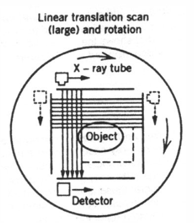
From the diagram, you can see clearly that the first generation has only one source, one detector and a single x-ray beam. The object is an example of a patient who needs a brain scan. This means a long linear translation movement before it can rotate. Scans with this generation of CT took a few hours and as we already knew that this is not good for human body because prolonged exposure can result in too much absorbed radiation.
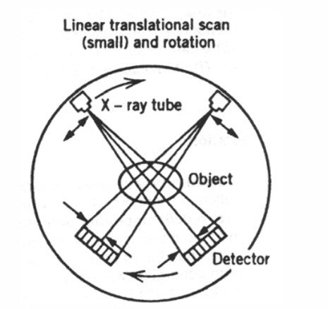
You can see from the picture that second generation added one more detector. And thanks to the additional detector, with more targeted scanning with a short linear translation movement and rotation, it helps to shorten the scanning time and hence, the radiation exposure to the patient. Is this good enough? No!
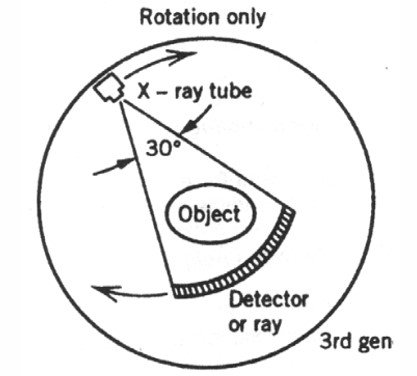
Here comes the 3rd generation of CT. This uses one source of x-rays with fan-beam and a row of detectors long enough to capture whole measuring range. This means that the machine only needs to do rotation and no more time-consuming linear translation movement.

For the 4th generation, it is inherently a similar design but the detector rows are now on the tube interior. In this design, this means the detector don't have to rotate anymore. Only the tubes needs to rotate.
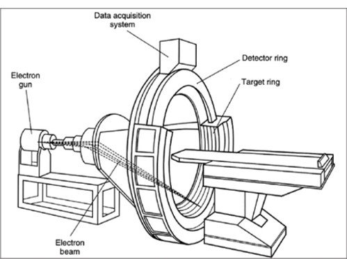
In this fifth generation CT, instead of using x-ray, the classical tube uses electron beam which acts as the cathode and the detector ring as the anode. When the detector ring is hit by the electron stream, it produces x-rays on the object. This means no more moving parts at all in this design and scanning can take place at a faster and more precise rates. In fact, there are more generations of CT machines and I shall leave it to @ct-gurus to talk more about it in future!
If you would like to connect to @ct-gurus for business, feel free to get in touch with them on steemit! They will be writing on CT (of course!), MRI and Biomedical Engineering on steemit. I wish all the best to @ct-gurus and look forward to their future blog entries. So follow @ct-gurus if you are interested to learn more on CT! Half of steam dollars from this post will go to @ct-gurus for thanking their contribution to steemit #academia #science.
We hope that you have learnt something from this post. We also welcome any feedback on #academiaspotlight. Please let us know if you found posts which are science and academic in nature which have not received as much love and we may add them to the next issue.
Stay tuned and watch out for #academiaspotlight issue 5!
You do such a great job culling through the science posts and finding completely ignored well written posts by knowledgeable posters. I have followed @ct-gurus and will look more into his posts.
Thanks! :-) i am going to dig more past gems and hopefully, they are still around..
I was teaching such lab classes about 10-12 years ago. The experimental platform was very similar to the one of second generation introduced above.
Aaaah memories memories ;)
wow... And i have never seen an actual one, not even experimental... only by a little theory...
It is actually pretty small (at least the one we had for the lab classes), and you do not need much to build it (although I didn't build it myself).
It was a round table with detectors that could rotate around its circumference. Sources were put in the middle of the table, hidden (at least for the students). By rotating the detectors, they were supposed to localize precisely the sources.
Nice. So many ways we can educate the students about the basic science principles...
I have experimented with something similar, DXA scanner, for personal body composition measurements. with DXA radiation is much lower and the practice is inexpensive.
Interesting! many applications using the same principal :-) saw the DXA wiki https://en.wikipedia.org/wiki/Dual-energy_X-ray_absorptiometry seems that it looks more quantifiable.
I wonder what is it with the loud humming noise that requires patients to need headphones to minimise it...