Ultrafast ultrasound imaging
Disclaimer: Background information
- In order to learn more about conventional B-mode sonography, please check out this article. It might help you to get a grasp of the underlying technology.
- This article aims to provide a gentle introduction into ultrafast ultrasound imaging. If anything is unclear or if there are too many confusing details, please let me know in the comments. I tried to keep some distracting details out of this post, while conserving the general message of this article.
Introduction
Conventional B-mode imaging has many advantages including relative ease of implementation and relatively low hardware requirements. However, it cannot be used for advanced ultrasound imaging modes like transient elastography, ultrafast vector tissue Doppler imaging or shear wave elastography. This is due to the low imaging frame rate of less than 100 Hz. For the advanced modes mentioned above, a relatively recent technology called Ultrafast Imaging is needed [1].
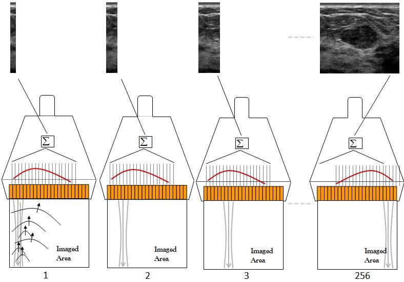
Conventional ultrasound imaging (Source: [2])
In contrast to conventional ultrasound image acquisition (as depicted in the figure above), ultrafast imaging is an inherently parallel technique which is capable of visualizing a full ultrasound image from one single ultrasound insonification, regardless of the size and the characteristics of the image. Thus, the imaging frame rate for ultrafast imaging is not limited by the number of reconstructed lines but by the time a single plane wave needs to propagate in the medium and to get back to the transducer again [2]. Such a plane wave is generated by activating all transmit elements of a linear array ultrasonic transducer at the same time as shown below.
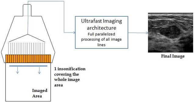
Ultrafast image acquisition (Source: [2])
Achievable frame rates
The table below compares the achievable frame rates (in Hz) for conventional ultrasound imaging and ultrafast imaging. It is important to note that ultrafast imaging requires sophisticated hardware to transfer and process the huge amount of data generated during the acquisition. Currently, the processing of the enormous amount of resulting data can not be done with a normal CPU in real-time but has to be done with special Field-programmable gate arrays (FPGAs) or GPUs instead.
| Application | Typical imaging depth | Conventional architecture (maximum frame rate) | Ultrafast architecture (maximum frame rate) |
|---|---|---|---|
| Abdominal imaging | 20 cm | 20 Hz | 3800 Hz |
| Cardiac Imaging | 15 cm | 150 Hz | 5000 Hz |
| Breast imaging | 5 cm | 60 Hz | 15000 Hz |
Comparison between conventional ultrasound imaging and ultrafast ultrasound imaging (Source: [2])
Image quality and plane wave compounding
Even though plane wave imaging allows for imaging at very high frame rates, the image quality in terms of contrast and resolution is much lower for ultrafast imaging compared to conventional ultrasound imaging. The reason for this loss of quality is the fact that the transmit focalization step is removed [1]. In the figure below the extent of this problem can be seen if the image taken at 25 Hz on the left hand side is compared to the picture taken at 18,000 Hz with ultrafast imaging.
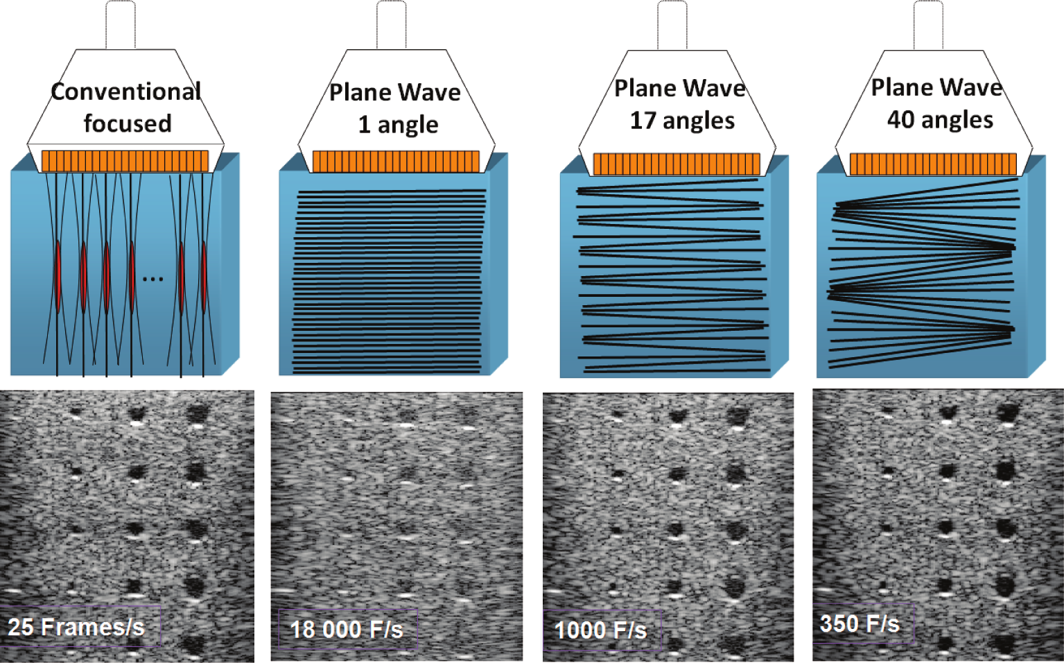
Comparison of conventional focused (leftmost image) and ultrafast ultrasound imaging using different angles (images on the right) (Source: [1])
The conventional focused ultrasound image taken at 25 Hz achieves a much higher contrast than the ultrafast image. Fortunately, a remedy to this problem exists as one can prevent loss of quality significantly by using different angles and creating a compounded image. The more different angles are used, the better the image quality gets, resulting in an ultrafast image having comparable contrast to a conventional image if the number of different angles is chosen to be high enough. The figure above (on the right hand side) shows how more angles (17 and 40 in this example) impact the image quality and imaging frame rate (1000 Hz and 350 Hz, respectively). It can be clearly seen that the maximum achievable frame rate drops proportionally to the amount of different angles used in order to obtain the image.
Effects of having more different angles
The graph below shows the effect the amount of different angles used for plane wave insonifications has on the contrast of the resulting image, measured in dB (lower values means higher contrast). The conventional 4-focal multifocus ultrasound technique, changing the respective foci between 1.1 mm in a focal plane to 1.6 mm at equal distance of 2 focal planes, is compared to plane wave ultrafast ultrasound imaging [3]. One can see that with 45 angles, the same contrast as with the 4 plane multifocal method is achieved. More than 45 angles can even lead to higher contrasts than conventional B-mode imaging.
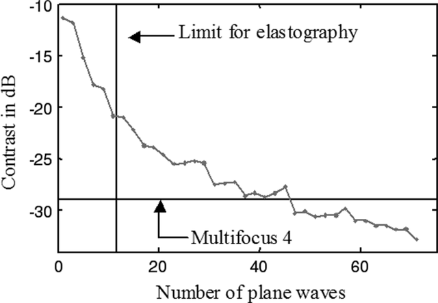
Effect of the amount of angles on image contrast for plane wave imaging (Source: [3])
Angle compounding in more detail
The minimum frequency of the shear waves used in elastographic imaging is 500 Hz [2] and thus the displacement needs to be imaged at (at least) 1 kHz to obey the Nyquist limit. In a future post we might learn more about the Nyquist limit and its importance but for the moment I don't want this article to become too long and cumbersome to read.
More than 12 different angles lead to imaging frame rates which are too low to be feasible for functional ultrasound imaging methods (like shear wave elastography). The figure below shows how the compounding is applied for three angles.
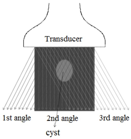
Applying angle compounding for three angles (Source: 4)
In diagnostic imaging the usage of several different angles is very crucial as the user usually would like to get high quality results. However, for functional imaging (e.g. imaging relying on the measurement of shear wave velocities such as shear wave elastography) it is not necessarily indicated to use different angles. In many cases, the measured effects can be analysed using just one single angle and only one insonification of the region of interest.
Higher frame rates during image acquisition allow for measuring effects which were impossible to observe previously. One example would be the possibility to visualize the propagation of shear waves by measuring the displacement induced by those waves. A future article might provide further details about this effect and its applications...
References
[1]: M. Tanter and M. Fink. “Ultrafast imaging in biomedical ultrasound”. In: Ultrasonics, Ferroelectrics, and Frequency Control, IEEE Transactions on 61.1 (Jan. 2014), pp. 102–119. ISSN : 0885-3010. DOI : 10.1109/TUFFC.2014.2882.
[2]: Jeremy Bercoff. “Ultrafast Ultrasound Imaging, Ultrasound Imaging - Medical Applications, Prof. Oleg Minin (Ed.)” In: (2011). URL : http://www.intechopen.com/books/ultrasound-imaging-medical-applications/ultrafast-ultrasound-imaging.
[3]: Gabriel Montaldo et al. “Coherent plane-wave compounding for very high frame rate ultrasonography and transient elastography”. In: Ultrasonics, Ferroelectrics, and Frequency Control, IEEE Transactions on 56.3 (2009), pp. 489–506.
Interesting to learn about it. Lately I've been using USG so often. The doctor had earned a lot from me 😁😅
Thank you very much for your comment, @kobold-djawa. Ultrasound is a very useful tool indeed. Especially for pregnancies it's a perfect fit due to the non-invasive and non-ionizing technology. All the best for you. :-)
Excellent article!
Thank you very much for your comment and for resteeming the article. Much appreciated. :-)
really helpful and informative
Thank you but somehow I really doubt that you read the whole article in 54 seconds. ;-)
lol I'm still reading it, but wanted to comment before I forget :)
I see... Thanks a lot for your comment and don't let any further comments interfere with the concentration you need to finish reading the post above.
the article was really nice being a student I find this helpful and interesting. I like the fact that you explained it well
Thank you very much for your feedback. Just let me know if there are any remaining questions. Good luck with your studies!