Human Anatomy 101 - The Eye (Detailed)
G'day team,
This article will require a working understanding of anatomical terminology. If you haven't read my posts on understanding anatomical terminology you can find them here
Anatomy 101 - Anatomical Terminology (Basics)
Anatomy 101 - Anatomical Terminology (Detailed)
If you're interested in a more basic overview of eye anatomy please read this article first!!
Human Anatomy 101 - The Eye (Basics)
If you're ready to jump into the deep end then read on!
In the Basic article we covered some of the reasons the eye is so awesome, so I guess we'll just get stuck into it!
The Orbit
The orbit is the bony cavity of the skull that holds the eye, it's muscles and other associated structures. The orbit actually gives a great insight into just how complex the structure of the skull is, being somewhat of a junction for many of the bones that make up our facial skeleton and cranial vault.
These bones are labelled in the image below!
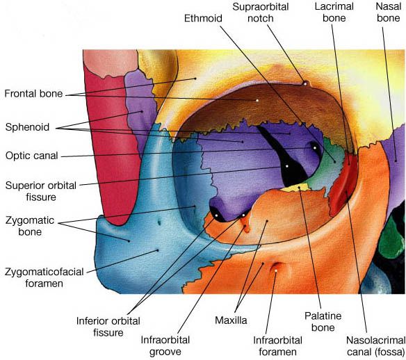
Image source
It's important to note the relationship of the orbit to each of these bones when considering facial trauma in patients.
Optic canal
This is a single hole you can see in the back of the orbit, medially and superior to the other holes. It's through this hole that the optic nerve and retinal artery access the eye.
Superior orbital fissure
This is the superior of the two long holes through the back of the orbit, the shape of which gives it its name 'fissure'. A number of important vessels pass through the superior orbital fissure, including...
The occulomotor nerve - responsible for moving the eye
Trochlear nerve - innervates ONLY the superior oblique muscle of the eye
Branches of the facial aspect of the ophthalmic nerve - innervates the upper face
Abducens nerve - innervates ONLY the lateral rectus muscle of the eye
Superior branch of the ophthalmic vein
Branches of the cavernus plexus
While it may seem like a memory game, understanding which vessels leave the cranial vault from which hole is extremely important. It means that when we find certain patterns of nerural deficit we can pinpoint which structures are involved
Inferior orbital fissure
This is actually a continuation of the long hole that forms the superior orbital fissure, but appears at a different angle and transmits only the zygomatic branch of the maxillary nerve and some smaller nerves.
Visual Axis
It's also important to note that from above the axis of the orbits (the directions they point) is not actually straightforward, but outwards at an angle. This means that while we sit here facing forward our eyes are actually both rotated inwards, so as to be focusing on the same object.
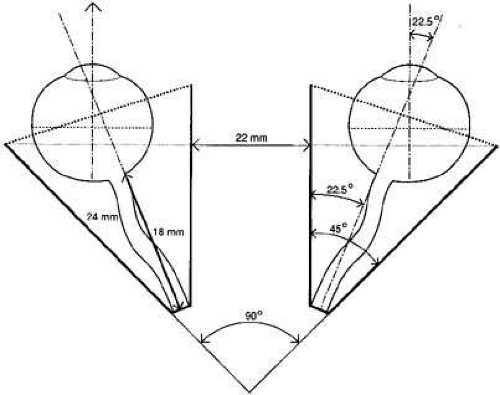
Image source
Now we understand the cavity that they eyes sits in, it's time to have a look at the muscles that surround, and move, the eye.
Muscles of the Eye
Yayyyy muscles! Everyone loves learning about muscles.
Let me change your mind : )
There are six extrinsic (outside) muscles of the eye, which are responsible for moving the eye. These include four straight muscles, and two which attach to the eye at an angle. Let's start with the strait ones:
Superior rectus
Inferior rectus
Lateral rectus
Medial rectus
These muscles all attach to a single fibrous ring sitting at the back of the orbit, and reach forward attaching directly onto the eye.
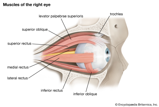
Image source
Each of these muscles is fairly simple. Each pulls the eye in a single direction. But there are two other muscles of the eyes, which are responsible for complex movements which we'll skip over here, but address in the detailed articles.
Superior oblique
The superior oblique muscle starts with the rest of the rectus muscles at the back of the orbit, it reaches forward running superior and medial to the eye (on the inside), it passes over the eye before attaching to a ligamentous loop called the trochlear, before turning around and passing back again to attach to the (here we go!) lateral, superior and posterior aspect of the eye. Complicated? Perhaps a diagram will explain it better!
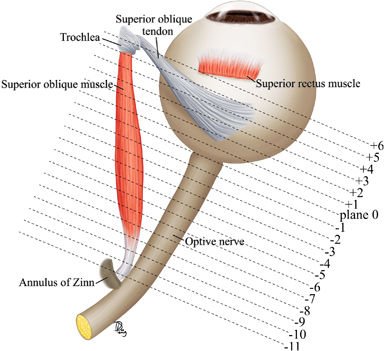
Image source - This is a RIGHT eye, as seen from above
Inferior oblique
Thankfully the inferior oblique is nowhere near as complex! It's the one extrinsic muscle of the eye that starts in front of the eye, medially, and reaches backwards laterally, attaching to the inferior, lateral and posterior aspect of the eye.
Innervation and Movement of the Muscles of the Eye
Now we understand the muscles themselves, let's have a look at how they work!
*The innervation to the muscles of the eye all comes from three cranial nerves. And they're conveniently named so it's super easy to remember...
Trochlear nerve - innervates the superior oblique muscle (the one which wraps around a trochlea
Abducens nerve - innervates the lateral rectus muscle (the one which abducts the eye)
Occulomotor nerve - does ALL the rest!
Movement of the eye is relatively simple if we remember one thing, the natural position of the eye if it is in line with the orbit is away from the midline of the body about twenty five degrees. If we remember that the eye and it's straight (rectus) muscles all work along this line then the rest is easy. So! The rectus muscles all pull the eye straight, relative to the orbital axis.
Superior rectus - elevation of the eye
Inferior rectus - depression of the eye
Lateral rectus - abduction of the eye
Medial rectus - adduction of the eye
Now, the oblique muscles. If you look at the diagram above you'll note that both oblique muscles pulls at it's attachment point on the eye from IN FRONT and MEDIALLY. This means they're pretty useless on an eye in a normal position, but if we adduct the eye, all of a sudden they become the elevators and depressors.
Superior oblique - depression of an adducted eye
Inferior oblique - elevation of an adducted eye
Now we understand the muscles that surround and move the eye, we should have a look at the vessels that enter the eye
Vessels of the Eye
There are many important vessels (arteries, veins and nerves) that enter the orbit and supply either the eye or the face, but seeing as we're just talking about the eye today we'll only cover the vessels related to it.
Ophthalmic Artery
The ophthalmic artery branches off the internal carotid artery, before it goes on to form the circle of willis, and enters the eye through the afformentioned optic canal. Right after entering the orbit it branches into a number of smaller arteries, the most important of which is the retinal artery. The retinal artery enters the sheath that surrounds the retinal nerve and goes forward in to the eye where is supplies (you guessed it) the retina. The other branches of the ophthalmic artery supply the muscles of the orbit and of the eyelids and face.
Optic Nerve
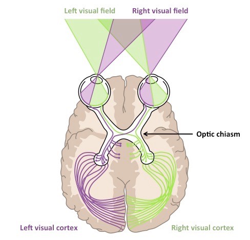
Starting at the eye the optic nerve passes backwards through the optic canal and continues backwards sitting right up under the brain before reaching the optic chiasm, it's here that the half nerve tracts decussate (switch sides). This occurs in a confusing but rational way. Let's talk through it. We know all the visual information from the right is processed on the right side of the brain. Both eyes recieve information from the right side of the body, and in both eyes it is recorded on the left side of the retina. This information travels along the left side of the left optic nerve (on the outside) and DOES NOT cross over at the optic chiasm, moving on to be processed on the left side of the brain. Now information on the left side of the body hits the left retinal field of both eyes. Let's look at the left eye. The left half of the retina feeds into the left half of the optic nerve and when it reaches the optic chiasm, crosses over on to the right optic tract and goes on to the right side of the brain. It is this way (by crossing over only information from the medial retinal fields or lateral visual fields) that information fromt he right side of the body ends up on the left side of the brain and visa versa. After the optic chiasm the optic nerve becomes the optic canal and enters the brain.
And finally, now we understand everything that surrounds it, let's look at the eye itself!
The Eye
Despite the complexity of it's function the anatomy of the eye is based on a very simple model. In essence the eye has just three layers.
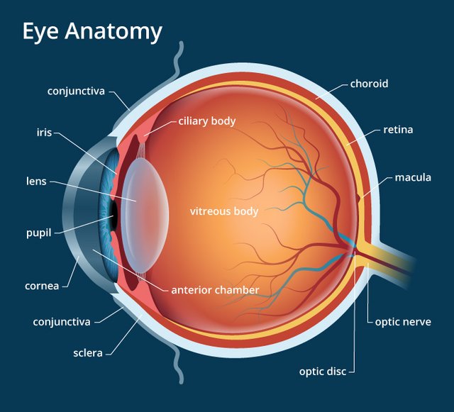
Image source
Sclera and Cornea
This is the outermost, protective, layer of the eye . It surrounds the entire eye and provides protection from insult. Over the lens, where light must pass through, it bulges out slightly and becomes known as the cornea. While the lens is responsible for fine-tuning the focus of our eyes, most of the actual bending of light occurs at the retina.
Choroid,
This is the layer of the eye that carries both the blood supply and the muscles. Anteriorly this includes the ciliary body, the muscles responsible for changing the shape of the lense when we focus, and the muscles of the iris which change how much light enters the eye. The ciliary muscles have an anchor-like attachment to both the eye and the lends, the result of this is that muscles lets the lens relax, while relaxation leads to contraction of the lens.
Retina
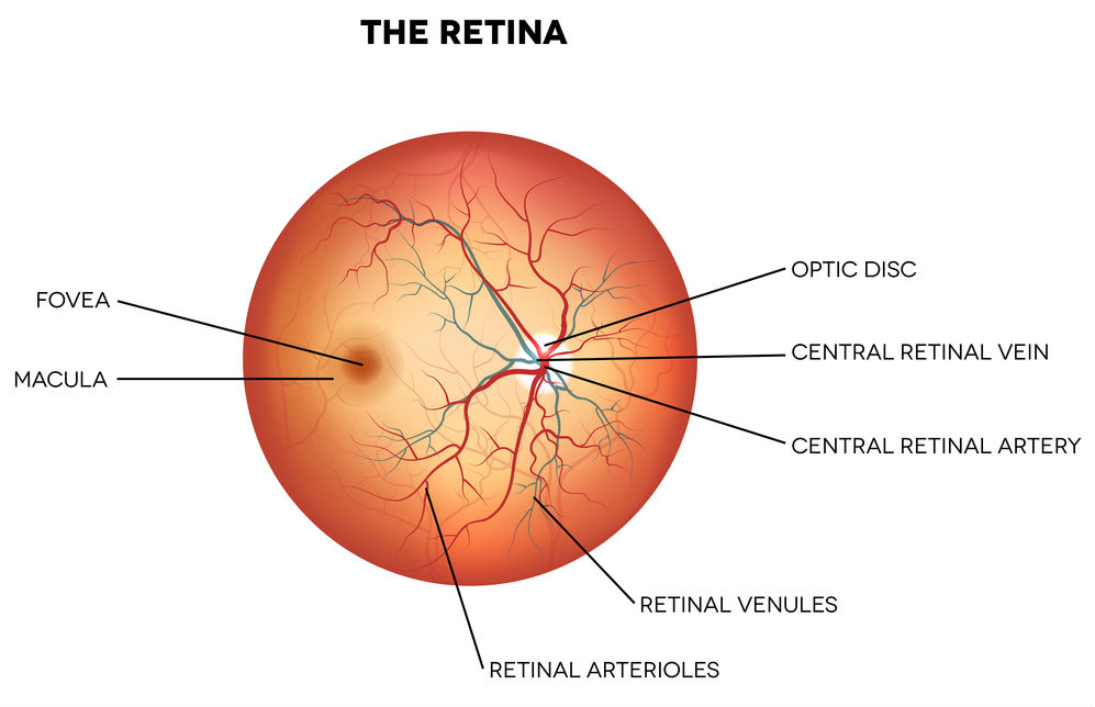
This is where all the fancy conversion of light into nerve signals occurs, this is the neural layer that covers the back portion of the eye. There are a number of regions on the retina that are important to note. The first is the macula, which is the centre of the retina and the section that forms our central vision and is particularly sensitive to colour images. At the very center of the macula is the fovea centralis, a small pit which is responsible for the high actuity at the very center of our vision.
The second is the site where the optic nerve leaves the eye, a small section which is not covered in retina and as such a small section at which we are blind. This 'blind spot' can be easily identified with a simple test.
Image processing in the retina may not be strictly anatomical feature, but it is fascinating none the less. The imaging processing in the retina occurs in the opposite direction to the light entering the eye. A pigmented layer at the back of the retina traps light that passes through nerve cells and prevents it from reflecting back into the eye. Cone and rod cells at the back of the eye react to being hit by light and feed information forward to cells known as horizontal cells, these in turn signal amacrine cells and these in turn pass information on to ganglion cells which converge and finally form the optic canal.*
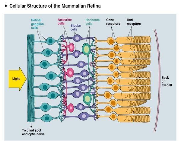
Image source not available
Eyelids
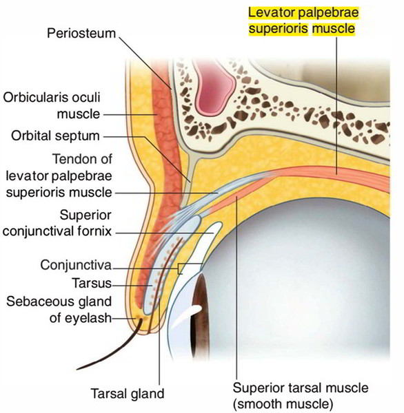
Now we're 90% of the way there, but I promised your information on the anatomy of the eyelids, and while this might seem a rather trivial (read pointless) part of the body to know in detail, it's actually very important. The eyelids recieve neural innervation froma number of nerves, and dropping of an eyelid (either partial or full) can indicate damage to either these nerve, the regions around these nerves or the hormones in the body that control the activity of these nerves. So let's get started.
There are two main muscles that hold the eye open
Levator palpabrae superioris
This muscle sits in the orbit with the rest of the extraocular muscles, innervated by the occulomotor nerve.
Superior tarsal msucle AKA Muller's muscle
This is an often forgotten little muscle that attaches to the tissue above the eyelid and then joins with the levator palpabrae superioris in the eyelid, it is a smooth muscle and innervated by the sympathetic nervous system, specifically fibers which origionate from the superior cervical ganglion at the top of the chest.
And one which holds it shut
Orbicularis Oculi
A very boring muscle which sits around the eye and helps squeeze is shut
That's it!
That's it, you've completed the anatomy of the eyes! Feel free to pop by next time for more anatomy fun :)
Thanks team
-tfc
My Recent Posts
Hi, I'm Tom! (My life of medicine, science and fantasy)
Super Bugs - What is antibiotic resistance and how we are fighting it
Hydatidiform Mole – When Pregnancy Goes Wrong
Tags - Top 20 Profitable, Engaging and Popular tags
New Years Resolutions - How to set goals, and achieve them
Great article,
Would mind adding your references for the text please:)
This post might help you understand why I am requesting for the references to be added: https://steemit.com/steemstem/@steemstem/being-a-member-of-the-steemstem-community
You can also join the discord chatroom: https://discord.gg/93dXBG5
Thank you:)
Ahh busted! No you're right I should be referencing, my main issue is 90% of this comes from memory, and the bits I look up come from a textbook. That being said I'll add something mentioning the texts and try and find an online backup for my work too!
Thanks :)
It's a sooner or later anyway. I gave you a vote, because I know that in time your content will get better. It should worth more, i know.
Good post, though I'll admit I'm a little squeamish with eyes. I hate it when the doctor is like, "I'm going to blow some air at your eye" because I can't close it, lol.
The eye is quite complex - great read.
Yeah, the corneal reflex is one of the harder to suppress reflexes in the body. Fun fact, a noise over 80db can also illicit it. Try this out by snapping your fingers in someones ears, their eyes will flicker :p
good job!
Thanks :)