How can our heart keep beating on it's own, over and over till we die - Understanding Electrical Activity of the Heart
Have you seen those horror movies where some one gets their heart ripped out from their chest and while the ghost/vampire/werewolf is waiting to eat the heart, the heart is still beating in it's hand? Or those cute gifs where some one is taking their heart out to give to their loved ones, still beating?
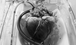
Well, as you can see, it's not entirely fiction. Given the proper nutritional environmental conditions, the heart can spontaneously keep beating on it's own even after being taken out from the body. Isn't it absolutely wonderful?!!
Today I'll try to explain you why is it that the heart can keep beating on it's own.
Exactly what is the heart? In simplest words, it's muscle. A muscular organ made of cardiac muscle cells. and what do muscles do, they contract and relax, they contract and relax...!! Contraction and relaxation of skeletal muscles creates physical movements that helps our body to physically move around.
Contraction and relaxation of cardiac muscles, instead of creating physical movement, moves blood around the body. I'll do a more detailed post on the cardiac cycle later in another post, but just to give a general idea, when the two ventricles of the heart contract, it pumps blood out from the heart into the lungs (from the right ventricle) and to the rest of the body (from the left ventricle). When the ventricles relax, they're basically filling themselves up for the next contraction.
Now, if you've been following my blog, you already know that an Action Potential has to be generated and transmitted to the muscle cells to create changes within the cell to cause the muscle contraction.
One important property to know about cardiac muscles is that, they are electrically connected, via something called Gap Junctions. What this means is, once an impulse has been generated, it will rapidly (almost immediately spread to all the connected cells so that they all contract at the same time. Once an impulse is generated, the two atria will contract at the same time, and then once the impulse is transmitted to the ventricles, the two ventricles will contract at the same time. This property is known as an electrical syncytium.
When we spoke about skeletal muscles, the impulses came from neurons, which resulted in contractions right? In fact almost everything in the body receives their impulse from nerves. If you have a stroke that damages the neurons that supply the muscles of you left leg, your left leg is paralyzed. If the damage was to nerves associated with sensation, you lose sensation in a particular area.
The twist
But how is it that when we take the heart out of the body, in the absence of all neuronal connections, the heart is still able to keep beating? Very good if this question popped up in your head!!
The reason is the heart doesn't rely on neuronal control from the brain to generate it's action potential. But this doesn't mean that in our body the heart doesn't have any neuronal connections at all. the heart does have neuronal connections with the brain, but those are used to control how fast or slow the heart should beat, or how strongly the cardiac muscles should contract. These neuronal connections do not generate the action potential, they only modify it. for example, when you exercise, your brain detects the need to supply more blood and nutrition to your body, and it will use it's neuronal connections with the heart to make it beat faster. But the heart was still beating before the brain asked it to beat faster.
So where is the the action potential/impulse for heart muscles generated? On the right atrium of the heart there is a specialized group of cells called Sino Atrial Node. The name comes from it's location. The atrial part you already get, it's in the atrium, duh!! But exactly where in the atrium? It is located near the opening of the superior vena cava, in a region called the sinus venosus.

When you discussed about action potentials, we saw after the action potential has been generated and has run it's course, the membrane potential eventually went back to the resting membrane potential and stayed there till another strong enough impulse came to depolarize the cell to it's threshold.
But what's so special about the SAN that it can keep generating impulse to take the membrane potential to the threshold and cause muscle contraction, over and over and over and over again from the moment your heart started functioning, till the moment you die!!!
Before we look at the critical difference in SAN, let's just quickly look at the action potential that passes through the rest of the heart muscle (of course after the impulse is generated from the SAN).

image from Kaplan USMLE Step 1 Physiology Lecture Notes
This is almost similar to the action potential we saw in the nerves. Let's look at a picture from that post again.

You see, the first part, the depolarization part is the same. The way this depolarization is created is also the same. Do you still remember how the depolarization happened in the nerve? Yes, influx of sodium via fast sodium channels!! (Feel free to brush up on what it means by fast and slow channels from the previous post, these terms are going to be used).
What we saw in the nerve cell is, once the repolarization starts, the membrane potential falls straight back. But in most of the cardiac muscle cells this part is slightly different. The reason being as the repolarization starts due to the fast sodium channels are closing, and some potassium is exiting via ungated channels, calcium channels open for a short while allowing Ca2+ to go in. This influx of +ve charges counteracts the fall in membrane potential and slightly prolongs the duration of the action potential.
Relax, slow down a bit mate
This is important in heart muscle because we need the ventricles and atria to contract long enough and squeeze out more blood from these chambers. Take a ketchup bottle at a restaurant for example. The amount of ketchup you'll be able to squeeze out if you squeeze the bottle very quickly and release immediately will obviously be much less than what you could squeeze if you squeezed for a little longer, right?!!

After the calcium channels close, the repolarization continues in the normal fashion as we've already learned. But something very interesting happens with the action potential of SAN. Let's see if you guys can spot it!!

image from Kaplan USMLE Step 1 Physiology Lecture Notes
Anyone? No?
See how there is NO Resting Membrane Potential?
Well no stable resting potential at least!! There is no flat line in the action potential. If you noticed in the rest of the cardiac muscle, phase 4 was a flat line. That was their resting membrane potential. Once it reached there, unless they were disturbed, the would stay there. Wouldn't depolarize. But look at how the phase 4 of the SAN looks like. It's not a flat line. As soon the repolarization happens and the potential reached the resting potential , the membrane potential automatically started to move up towards the threshold and once it reaches, an action potential is generated and the cycle keeps repeating itself from the moment your heart started functioning in your mother's womb till you die!! How wonderful is that!!
So what is making SAN automatically depolarize itself?
The SAN has something called the "funny current" going on!! Recall that in almost every other action potential, during the resting membrane potential, the sodium channels are closed and there is little to no sodium entering the cell. The voltage gated sodium channels open when the cells depolarize and influx of sodium marks the beginning of an action potential, and these channels are closed when the cell repolarizes.
These sodium channels are also voltage gated, but they are "funny" in the sense that they open when the cell repolarizes and close when depolarization begins. So while almost every other cell, on repolarizing, takes a break till the next potential comes, the SAN cells start slowly depolarizing themselves automatically as soon as the repolarization happens, because repolarization triggers the opening of these funny voltage gated sodium channels!!
These channels are not as fast as the ones we previously encountered. The influx of the sodium ions occur relatively slowly through these channels which is why you see the membrane potential rising slowly towards the threshold, instead of the rapid upstroke we saw with sodium influx during action potential.
There is one more small difference that is good to know (not absolutely necessary right now, but will be slightly importance in future posts) and then i'll wrap up this post!!
So after the depolarizing and reaching threshold, these funny sodium channels will close. Then how does the upstroke of action potential happens for SAN cells? Unlike what we've seen, the upstroke here happens due to opening of slow calcium channels and a slow influx of Ca2+ ions. You'll notice that the upstroke of SAN cell action potential is not as steep and quick as we've seen before. This is becasue so far we've seen depolarization via fast acting channels and rapid Na+ influx. But here, we have slow acting channels and a slow Ca2+ influx.
Sources :
Kaplan USMLE Step 1 Physiology Lecture Notes, 2013
Guyton and Hall Textbook of Medical Physiology, Twelfth edition
Cardiac Action Potential
Phases of cardiac action potential

If you enjoy medical topics, or health tips, please make sure to follow me at @simplifylife
Peace!!
Quite a perfect organ, and yet one of the main causes of death. Hopefully we'll soon either replace them with machine hearts, or else find a way to rejuvenate them.
Good reminder that it is the electrolytes in our bodies that are responsible for our heart contractions and rhythm. I have seen patients develop atrial fibrillation from being on hydrochlorothiazide for too long without a proper diet.
Also try centering your pictures. all you have to do is surround the full picture link with (center)picture link(/center) but use <> instead of ().
Thanks so much for taking your time to read the post :) Really appreciate it.
Yeah it's really important to counsel the patients to maintain a proper diet when prescribing diuretics. And Thiazide group is specifically know for it's side effect of hypercalcemia, and with calcium playing big role in the electrical activity in the heart, it's not surprising why thiazides can disrupt normal rhythm.
Also, thanks a lot for the code. Also, do you happen to know how to take pictures on either side of text? I wanted to try that too with some posts but didn't know how.
Once again, thank you very much.
I tried using the code, but it didn't work :(
There are a number of old posts on here that can likely show you how to do the coding properly better then what I can explain without having to take pictures. do a google search of markdown picture center justification and something should come up.
Gotta be careful with those potassium/calcium wasting diuretics--can lead to hypkalemia and hypocalcemia.
A great post and a great breakdown of the autorhythmicity of the heart! You do a great job of simplifying the whole process too so anyone could understand it!
Thanks so much for the kind words tom!! I try my best :)