Fluid and Electrolyte Imbalances: Electrolyte Deficits
Electrolyte Deficits

An electrolyte, as the name suggests, is the term that receives any substance capable of acting as a conductor of electricity. Knowing that our body works thanks to electrical impulses sent by neurons, we can infer the importance of electrolytes in our physiology, the most common of them in intravascular fluids being sodium, chlorine, potassium, and bicarbonate.
These substances can become scarce, causing pathological disorders that can be life-threatening. Electrolytic deficits are common, for example, in surgical patients; in which there is a loss or redistribution of extracellular fluid to nonfunctional spaces, such as the so-called third space (the space found between the cells, also called interstitial space), either due to trauma or ischemia during or before the operation, or because of diseases such as pancreatitis, peritoneal sepsis or intestinal occlusion. The electrolyte deficits are classified according to the electrolyte that is diminished, the most medically relevant are:
- Hyponatremia: It is defined as a plasmatic sodium concentration below 135 mmol / L, its main cause is an alteration of the renal excretion of loose water, due to an increased secretion of vasopressin (a hormone that increases the tonicity of blood vessels and decreases the volume of urine). This causes an excess of water in relation to the amount of sodium, decreasing its osmolarity. Hyponatremia has several classifications, the main ones are:
According to Sodium Concentration: it is divided in mild (130-134 mmol / L), moderate (125-129mmol / L), and severe (<125 mmol / L).
According to its Evolution Time: in acute hyponatremia (evolution time less than 48hrs) and chronic hyponatremia (evolution time longer 48hrs)
As for the clinical picture the signs and symptoms will depend on the amount of sodium still present in the blood. The main alterations are in cognitive functions and balance; nausea without vomiting, confusion, drowsiness, stupor, seizures and even coma. If the patient also presents polydipsia, skin and mucous dryness, tachycardia, and oliguria, the hyponatremia may be accompanied by isotonic dehydration, and it should always be suspected when the signs and symptoms appear after a surgical operation or intense physical effort. , or in patients who are being treated with diuretics; such as those who suffer from edema.
The diagnosis of this pathology, as in all electrolyte deficits, is quite simple: just measure the amount of plasma sodium and see if it is below the normal value of 135 mmol / L, and then do a differential diagnosis based on clinical signs with other diseases, such as hyperglycemia or psudohyponatremia. The treatment is also relatively simple; the goal is to replace the sodium lost with 0.9% or 3% saline solution, normally transfused at a rate of 100ml / hr, although this time varies according to the rapid evolution of hyponatremia; the longer the evolution, the slower the replacement of sodium should be, and vice versa. The amount of solution to be used can be calculated taking into account that there is commonly an increase of 10 mmol / L of sodium for each liter of solution administered, in order to avoid administering an excessive amount, the following formula is used:
The plasmatic sodium level the patient must be taken to is 130 mmol / L, we must also remember to stop the administration of oral fluids unless the patient also has dehydration, and seek to eliminate the cause of the hyponatremia if possible.
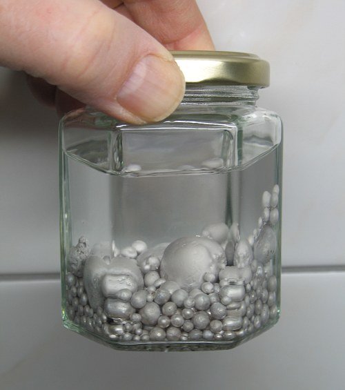
- Hypokalemia: It is defined as the decrease of blood potassium values below 3.8 mmol / L, it can be caused by base pathologies such as hyperaldosteronism, Gitelman syndrome, or Cushing syndrome , although most frequently it occurs because of an insufficient nutritional intake of potassium, an alkalosis, burns, or an excessive administration of diuretics or laxatives.
The patient will present manifestations related to the blockage of the action potential that occurs, such as dangerous arrhythmias, muscle weakness, constipation, urinary retention, paresthesias and in some cases severe rhabdomyolysis (necrosis and muscular dissolution). The severity of the condition depends on how much the potassium is decreased, and the speed at which it is lost. For its diagnosis, simply determine the potassium concentration in blood with laboratory studies, and check their values, confirming hypokalemia if it is below the normal value of 3.8 mmol / L.
The treatment is based on the administration of potassium in the form of Potassium Chloride (KCl), and of potassium-sparing drugs, such as Amiloride and Spironolactone, the latter being the first choice. The potassium deficit is repaired as follows:

- Hypocalcemia: It is the decrease in the bodily calcium concentration below its normal values of 2.25 mmol / L, or 9 mg / dl. Within its causes, the main one is a low intake of calcium in the diet, although it may also be due to there being enough calcium intake, but it accumulates excessively in soft tissues or bones due to various disorders such as pancreatitis, hyperphosphatemia , or after an extraction of the parathyroid gland. It can also be caused by alterations in the intestinal absorption of calcium, by its loss in urine due to diuretic consumption, or by deficits of vitamin D or parathyroid hormone.
The signs and symptoms are mainly of neuromuscular character, its main manifestation is tetany; involuntary muscular contractions that begin at the distal end of the extremities, progressing proximally, ending in the torso and face, in the form of labial contractions and on the eyelids. In addition to this characteristic symptom, there may be photophobia, diplopia (double vision), bronchospasms that can lead to an asthmatic crisis, angina pectoris, migraines, and a lengthening of the QT segment in the ECG.
For its diagnosis, besides being based on the decrease in the serum calcium concentration <2.25 mmol / L, it can be detected by two characteristic signs of the disease: the Chvostek sign, in which the facial nerve is tapped 2cm in front of the earlobe, and if positive, involuntary muscular contractions are seen in the lips, and the sign of Trousseau, in which a tensiometer is used and inflated 10 or 20mmHg above the patient´s normal systolic blood pressure levels for 3 minutes, and it is said to be positive when he begins to present contractions and pain in the hands.
As for the treatment, it is necessary to focus on eliminating the cause of the pathology, and then eliminate the deficit by administering calcium, according to the type of hypocalcemia; if the patient is symptomatic, 20 ml of 10% calcium gluconate is administered intravenously, repeating the dose each time the patient presents symptoms, while oral administration of calcium and vitamin D supplements begins. If it is a chronic hypocalcemia, calcium is administered from 1000 to 3000 mg per day orally, in the form of calcium carbonate or calcium acetate, as well as vitamin D in a dose of 0, 5 to 2 ng per day.
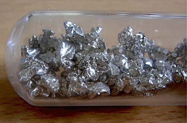
- Hypomagnesemia: This is the decrease of magnesium in blood below 0.65 mmol / L, caused normally by a low intake of magnesium in the diet, by alterations in its absorption in the digestive tract, renal magnesium losses (mainly due to tubulopathies), or by a mobilization of magnesium into the extracellular compartment, caused by the hungry bone syndrome (increase in bone uptake of phosphorus, magnesium and calcium), or by the treatment of diabetic ketoacidosis.
Its symptoms are quite vague and nonspecific, which makes its clinical detection quite difficult. It usually brings concomitant deficits of calcium and potassium that do not improve with supplementation, heart rhythm disorders of various types, tetany, tremor of the tongue, muscle weakness, and in some cases there may be a prolongation of the QT segment, and flattening of the U and T waves on the ECG. For the diagnosis, it is enough to perform a test of the blood magnesium concentration, and verify that it is below the normal values. It is necessary to remember to evaluate the values of the other electrolytes, since if there is hypomagnesemia, the other electrolytes are also usually low, as well as to determine the concentration of creatinine to detect possible tubulopathies.
To treat it, we seek to eliminate the causes of hypomagnesemia first. Then, we proceed depending on whether the patient has symptoms or not; if he is in a symptomatic state, 1 to 2 grams of magnesium sulphate are administered IV in 10 to 15 minutes, then in a slow infusion in a quantity of 5 grams diluted in 500 ml of 5% glucose solution for 5 hours; these amounts may increase according to the magnesium deficit. If the patient is asymptomatic, a magnesium preparation is given orally, taking into account that all these preparations cause diarrhea.
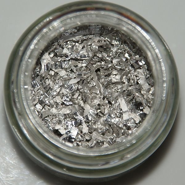
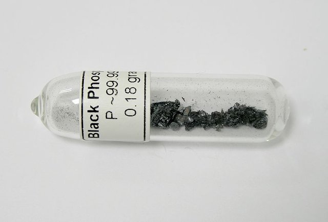
- Hypophosphatemia: It is a drop in the concentration of bodily inorganic phosphate below its normal values of 0.9 mmol / L. Its possible causes are very similar to those of a hypomagnesemia; low phosphate consumption at meals, poor intestinal absorption, renal phosphate losses due to hyperparathyroidism or tubulopathies, or mobilization of extracellular phosphate to the intracellular space by respiratory alkalosis or hungry bone syndrome. It is also common during the treatment of diabetic ketoacidosis.
Patients with hypophosphatemia will present mild symptoms at the beginning, limited to bone pain and muscle weakness. As the deficit worsens, symptoms such as involuntary muscle contractions, thrombocytopenic purpura, seizures that may progress to a coma, liver damage, and in severe cases rhabdomyolysis begins to appear. To confirm its diagnosis, one has to detect a decreased serum phosphate below the normal values, and the cause is determined by the evaluation of the other electrolytes, and complementary laboratory tests such as a urine test or a gasometry.
In the treatment, the cause must first be corrected, and then the phosphate deficit should be eliminated with a diet rich in phosphorus, and a mixture of 17.8 g of disodium phosphate, plus 4.8 g of sodium biphosphate diluted in 100 ml of distilled water daily, to be administered orally. This solution contains about 25-30 mmol of inorganic phosphors. In patients who do not tolerate the oral route, sodium phosphate or potassium phosphate is administered in amounts of 0.08 to 0.16 mmol of phosphorus per kg of the patient´s weight (taking into account that 1 ml of either of the two solutions has 2 mmol of phosphate) intravenously, remembering that a constant measurement of the inorganic phosphate and calcium concentrations of the patient must be maintained.
References:
- Armas, Rodolfo, et al. Internal Medicine Base on the Evidence 2017/2018, 2nd ed., 2017.
- Arias, Jaime, et al. Medical and Surgical Generalities, 1st ed., 2002.