!! The cell membranes ¡¡ ( Part 1/2)
To carry out the chemical reactions necessary in the maintenance of life, the cell needs to maintain an appropriate internal environment. This is possible because the cells are separated from the outside world by a limiting membrane, the plasma membrane. In addition, the presence of internal membranes in eukaryotic cells provides additional compartments that limit unique environments in which highly specific functions necessary for cell survival are carried out.
The plasma membrane is responsible for:
Selectively isolate the contents of the cell from the external environment
Regulate the exchange of substances between the cellular interior and exterior (what enters and leaves the cell
Intercellular communication
Most cells have internal membranes in addition to the plasma membrane, form and delimit compartments where the biochemical activities of the cell are carried out. The remaining membranes also constitute selective barriers for the passage of substances.
Membrane functions
The cell membrane functions as a semipermeable barrier, allowing the passage of few molecules and keeping most of the products produced within it.
Protection
Help subcellular compartmentalization
Regulate transport to and from the cell and subcellular domains
Serve receptors that recognize signals of certain molecules and transduce the signal to the cytoplasm.
Allow cellular recognition.
Provide anchoring sites for cytoskeletal filaments or components of the extracellular matrix which allows, among others, the maintenance of the cellular form
Serve as a stable site for enzymatic catalysis.
Provide "doors" that allow the passage through the membranes of different cells (gap junctions)
Regulate the fusion of the membrane with another membrane by means of specialized junctions
Allow address cell motility
Structure of the membranes
The plasma membrane has a thickness no greater than 5 nm. Because most of the proteins have a diameter greater than 10 nm, one of the main problems in understanding the basic structure of the membranes was to determine how the molecules were arranged in such a small space. The current model of the structure of the plasma membrane is the result of a long road that begins with the indirect observations that determined that the fat-soluble compounds easily passed this barrier which led Overton, in 1902, to maintain that its composition corresponded to the of a thin lipid layer; Subsequently, the proposal was made that the protein was also involved in the composition. Towards 1935 Danielli and Davson synthesized the knowledge proposing that the plasmatic membrane was formed by a "lipid bilayer" with proteins adhered to both sides of it.

The integration of chemical, physico-chemical data and various microscopy techniques led to the current model of "Singer SJ, and Nicolson, GL (1972) Science, 175: 120" (Singer SJ, and Nicolson, GL (1972) Science , 175: 120). According to this model of the fluid mosaic, which has had great acceptance, the membranes consist of a lipid bilayer (a double layer of lipids) in which various proteins are immersed.
The lipid bilayer has been established as the universal basis of the structure of the cell membrane. It is easy to observe in an electron micrograph but specialized techniques such as X-ray diffraction and cryofracture techniques are needed to reveal the details of your organization.
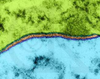
Opposed neuron cell membranes
Image source
The membrane is a quasi-fluid structure, in which its components can perform translatory movements within it. This fluidity implies that the components are mostly only joined by non-covalent bonds. The electron microscopy showed the plasma membrane as a structure of three layers, two of them external and dense, and one clear in the middle.
Lipids are insoluble in water but dissolve easily in organic solvents. They constitute approximately 50% of the mass of most of the plasma membranes of animal cells, with almost all the rest being proteins. There are 109 lipid molecules in the plasma membrane of a small animal cell.
The primary molecule of the cell membrane is the phospholipid, it has a polar "head" (hydrophilic) and two non-polar "tails" (hydrophobic), are therefore simultaneously hydrophilic and hydrophobic (Term introduced by Hartley in 1936).

Because molecules of the phospholipid type have one end that freely associates with water and another that does not, when they are dispersed in water they usually adopt a double layer conformation. The bilayer structure allows the groups of the hydrophilic end to associate freely with the aqueous medium, and the hydrophobic chains of fatty acids remain inside the structure, away from the water molecules.
Diagram of the fluid membrane model
- Bilayer of phospholipids)
- External side of the membrane
- Internal side of the membrane
- Intrinsic membrane protein
- Protein ion channel of the membrane
- Glycoprotein
- Phospholipid molecules organized in bilayer
- Cholesterol molecules
- Carbohydrate chains
- Glycolipids
- Polar (hydrophilic) region of the phospholipid molecule
- Hydrophobic region of the phospholipid molecule.