SENSES AND BEHAVIOUR: Photoreception in cone cells, Hearing, Balance, and Simple Behaviour.
Greetings to my ardent readers and to the entire steemstem community. This post is a sequel to my previous post on Senses and behaviour. In the final paragraph, I said there are two types of photoreceptor in the human eyes are the rods and cones, but I stopped at the explanation of photoreception in rod cells. Now, I will start this post by explaining the photoreception in cone cells.
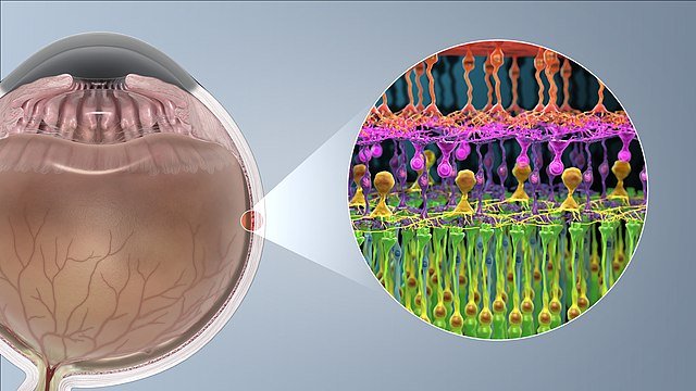
Photoreception in cone cells
The function of the cones is to see colour and
detail, although they can only do this in bright light. In dim light they do
not function at all. Cone cells contain the pigment iodopsin, a photosensitive
pigment made up of photopsin, a different protein from that found in rods, and
retinal. The events that occur in cone cells stimulated by light are basically
the same as those that occur in rod cells. There are, however, three different
types of cone.
Each one contains a slightly different pigment and responds to a different wavelength of light. One responds to red light, one to green light and one to blue light, so allowing us to see in colour.
The colour that we ‘see’, or more accurately perceive (interpret in our brain), depends on which cones are stimulated. When all the cones are fully stimulated we see ‘white’. When very few are stimulated we see black. Stimulation of separate types produces red, green or blue, and all colours in between are produced by combinations of different levels of stimulation of the three types together. This model of how three different types of cone can produce the range of colour vision that we experience is known as the trichromatic theory of colour vision.
A table comparing rods and cones
| Feature | Rod cells | Cone cells |
| Shape | Rod-shaped outer segment | cone-shaped outer segment |
| Connections | Many rods connect with one Bipolar neurone | Only a single cone cell per bipolar cell |
| Visual acuity low | Low | High |
| Visual pigment(s) | Contain rhodopsin (no colour vision) | Contain three types of iodopsin, responding to red, blue and green light |
| Frequency | 120 million per retina – 20 times more common than cones | 7 million per retina – 20 times less common than rods |
| Distribution | Found evenly all over retina | Found all over retina but much more concentrated in the centre, particularly the yellow spot or fovea centrals |
| Sensitivity | Sensitive to low light intensities (used in dim light) | Sensitive to high light intensities (used in bright light) |
| Overall function | vision in poor light | Colour vision and detailed vision in bright light |
Visual acuity: the ability to see detail
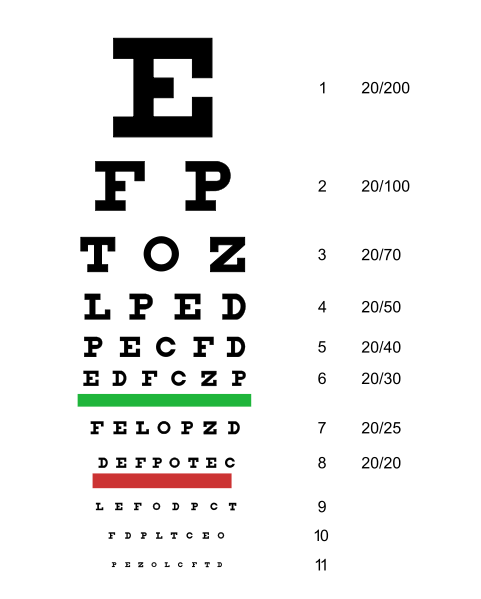
From what distance can you read a newspaper? Can you read the bottom line of the standard optician’s eye-test board? Tests such as these are a measure of visual acuity, which is defined as the ability to resolve detail. For instance, two lines such as these ====== will obviously appear as two lines, but if you tilt the laptop up and walk backwards, a point will be reached when they look like one grey line.
An interesting feature of visual acuity is that we only have a very narrow field of accurate vision. Focus on any particular word on this article and you see it in detail, but those on either side are unclear. We see only a very small proportion of our field of vision in detail – and that is the part that is focused on the fovea, and therefore that stimulates the cone cells.
Overall, rods are more sensitive than cones. Rods can function in poor light, but they cannot perceive detail; you can’t read small print in dim light. The cones have a higher visual acuity than the rods, but they need bright light. Why is there such a difference? The answer lies largely in a phenomenon known as retinal convergence.
Retinal convergence
There are over 120 million rods and about 7 million cones, but only about 1 million neurones in the optic nerve. Clearly, each rod and each cone cannot all have their own neurone. In fact, many rods converge into one neurone, while only a few cones are associated with each neurone. In the centre of the fovea it is thought that each cone actually has its own neurone,
The concept of retinal convergence generally is that, the further out you go from the centre of the fovea to the edge of the retina, the greater the degree of convergence. Two separate stimuli (such as two dots or lines) may stimulate two separate cones, but not the one in between. Therefore, the brain will see these as two separate images. The same two stimuli falling on the rods will not stimulate separate neurones, because a group of rods all feed into the same neurone. In this case, the brain will perceive only one stimulus, and see only one line or dot.
TRANSMISSION OF NERVE IMPULSES TO THE BRAIN
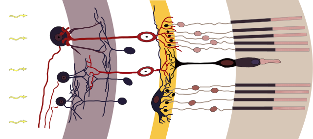
If you look back at the diagram of the photoreceptor cell, you can see that the photoreceptor cells are linked to a set of nerve cells in the retina called bipolar cells. These link with a second type of nerve cell called ganglion cells. It is the axons of ganglion cells that take information from the eyes to the brain. Ganglion cell fibres are bundled together to form the optic nerve.
We cannot see an object whose image falls on the retina at the point where axons of ganglion cells leave to form the optic nerve. Since it contains no receptor cells, any light striking this small area is not sensed; hence its name, the blind spot.
To demonstrate the blind spot, draw on a book two symbols such as a plus (+) and dot (.) evenly spaced out from each other. Then hold the book at arm’s length with the two symbols straight in front of your eyes. Close your left eye and concentrate on the cross with your right eye. Bring the book slowly towards you. Keep looking at the cross on the left: eventually, as the dot falls on the blind spot of your right retina, the image of the dot on the right will disappear
HOW THE BRAIN PERCEIVES AN IMAGE
Action potentials do not differ very much from one another and so there are only two possible ways in which stimuli going into the brain can be coded. One is the rate at which action potentials arrive and the other is their destination in the brain.
The rate at which sensory neurones fire tells the brain the strength of the stimulus: specific neurones tell the brain which part of the retina has been stimulated. The brain analyses this information to assess the nature of the original stimulus. This process, called visual processing, is immensely complex.
Specific areas of the cerebral cortex are associated with different sensory functions. The visual cortex in the occipital lobes deals with visual information. However, body senses must not be considered in isolation. Sensory and motor systems interact within the control regions of the CNS, and sensory information is processed at various points along the nerve pathway before it reaches the brain. In the brain it is processed further with information from other senses and stored memories.
HEARING AND BALANCE
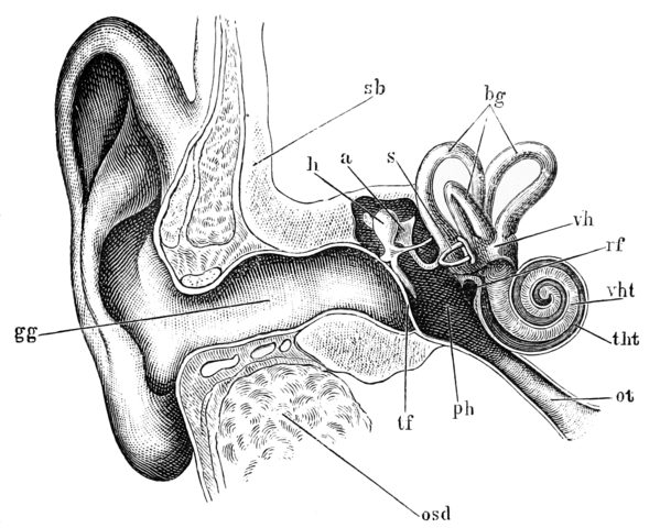
The ear is a miniature receiver, amplifier and signal-processing system. It is divided into three parts – an outer ear, middle ear and inner ear – and is connected to the brain by the auditory nerve. The sense of hearing that allows us to detect and process sounds, and the senses of balance and proprioreception that allow us to maintain our balance and posture, are possible because of the human ear. Sound waves pass through the outer and middle ear and into the inner ear through the oval window. Pressure on the oval window squashes the fluid in the inner ear, and compression waves travel through the canals of the cochlea. The round window eventually receives these movements of fluid and dampens them down (this prevents them from being reflected back into the cochlea) by bulging into the middle ear. Sensitive hair cells in the cochlea fire off signals, sending action potentials along the auditory nerve to the brain.
Schematic diagram of the human ear. Morten Bisgaard, Public Domain
BEHAVIOUR
SIMPLE BEHAVIOURS: One of the simplest types of behaviour is orientation. This enables organisms to move in order to find favourable environments and to avoid unfavourable ones. Animals such as insects show two types of simple orientation:
- Taxes: movements in a specific direction that are directed by a stimulus such as light or food.
- Kineses: more random movements that are not directed by a stimulus. Taxes and kineses are usually regarded as reflexes. A reflex is a rapid, automatic respose to a specific stimulus. It is a buit-in response: it does not have to be leaned. Reflexes also occur in higher animals, including mammals and humans.
Taxes
In many orientation behaviours, the animal moves directly towards or directly away from a stimulus. Maggots (fly larvae) have very simple eyes and can respond to the direction of light. Honey bees move towards a scent which is associated with food. These types of directional response are called taxes (singular: taxis)
A positive taxis involves movement towards a stimulus, a negative taxis involves movement away from the stimulus. As shown in the table below, taxes are also classified according to the type of stimulus involved (light, water, gravity) or to the type of behaviour shown (straight-line responses, turning movements).
Summary of some common taxes. These are normally classified either by the type of stimulus that triggers them or by the type of response they elicit.
| Name of taxis | Type of stimulus | Type of response |
| Telotaxis | Fixing on a stimulus without scanning from side to side | |
| Klinotaxis | Moving head repeatedly from left to right to judge stimulus intensity | |
| Geotaxis | Gravity | |
| Thermotaxis | Heat | |
| Phototaxis | Light | |
| Anemotaxis | Air currents | |
| Rheotaxis | Water currents | |
| Chemotaxis | Chemicals, e.g salts |
Kineses
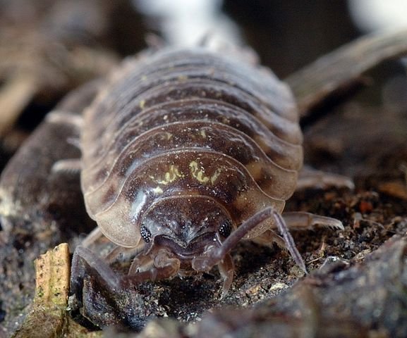
Woodlice are crustaceans, the group that includes crabs. Shrimps and lobsters. Woodlice are the only truly terrestrial crustaceans, but are prone to desiccation (drying out). Thus they need dark, damp conditions where they are also safe from predators and can find rotting vegetation to eat.
If placed in dry, light conditions, woodlice will move rapidly (by their standards), changing direction frequently. They don’t move in any particular direction, which is why this behaviour is classed as a kinesis. They can’t see or use any other senses to locate dark and damp places, but their behaviour means that sooner or later they have a good chance of finding the conditions they require. When they find dark, damp conditions, their movements are much reduced. Some common types of kineses are shown in the table below.
| Name of Kinesis | Type of stimulus | Type of response |
| Photokinesis | light intensity | |
| Chemokinesis | chemical gradients | |
| Hygrokinesis | humidity | |
| Klinokinesis | turns faster when stimulus is more intense | |
| Orthokinesis | moves with greater speed when stimulus is more intense |
MORE COMPLEX BEHAVIOURS
We can divide all animal behaviour into two categories: instinctive behaviour, or innate behaviour, which is inherited, and learned behaviour, which is modified by experience.
Instinct
There are many examples where a piece of behaviour appears without any previous experience. A herring gull chick automatically pecks at the red spot on its parent’s bill to get it to regurgitate food. The digger wasp, Ammophila, builds a nest in the sand and immobilises a caterpillar using precisely the night amount of poison injected into exactly the right spot, without any prior knowledge. We describe this sort of behaviour as instinctive.
Charles Darwin was the first to propose a definition of instinct in terms of animal behaviour. Natural selection, he argued, applies just as much to behaviour as it does to body parts and so behaviour can be inherited. Those animals that do not behave in the right way (e.g. finding the right food or looking for a mate) do not pass on their genes to the next generation.
But the classical, ethological view of instinct was put forward by Konrad Lorenz and Niko Tinbergen. They said that motivational energy – the drives and urges such as hunger, thirst and sex – control complex sequences of behaviour, which they called fixed action patterns. These behaviours, which include nest-building, courtship and parental behaviour, are automatic and occur in response to specific stimuli called releasers.
The concept of instinct has changed several times since the early ideas. Modern research has shown that instinct is not ‘driven from within’. Feedback from the body’s receptors continuously modifies behaviour associated with feeding, sex and grooming. Behaviours are often described as either instinctive or learned. But, in reality, the majority of behaviours rely on both inheritance and the effects of the environment.
Woodlouse activity decreases as humidity increases. André Karwath aka Aka, CC BY-SA 2.5
Learned behaviour
Learning is behaviour that is modified by experience. A 3-day-old chick eventually learns to peck at food, not at stones on the ground, and a bee learns to visit those flowers with most nectar. Such behaviours are useful because they allow animals to respond to changing conditions.
Some features of the environment, such as gravity and seasonal changes, are highly predictable. An animal does not need to learn how to respond to these situations: to do so would waste energy that could be used for other things. Animals learn:
- To avoid predators:
- To avoid harmful environments:
- To find food:
- To avoid harmful food;
- To find a suitable mate;
- To recognise important individuals within the group:
- To find their way home.
I will like to pause here for now. In my next post, I shall discuss more on learned response and complex learning.
Thanks for reading.
REFERENCES
https://www.britannica.com/science/photoreception/Structure-and-function-of-photoreceptors
https://en.wikipedia.org/wiki/Photoreceptor_cell
https://www.healthline.com/health/visual-acuity-test
https://en.wikipedia.org/wiki/Visual_acuity
https://www.sciencedirect.com/topics/psychology/visual-acuity
https://www.ncbi.nlm.nih.gov/pmc/articles/PMC1283714/
https://en.wikipedia.org/wiki/Retinal_summation
https://nba.uth.tmc.edu/neuroscience/m/s2/chapter14.html
https://medical-dictionary.thefreedictionary.com/retinal+convergence
https://www.dummies.com/education/science/understanding-the-transmission-of-nerve-impulses/
http://www.e-missions.net/cybersurgeons/?/nerv_teacher/
https://blogs.scientificamerican.com/mind-guest-blog/how-the-brain-processes-images/
https://science.howstuffworks.com/life/steady-view.htm
https://www.brainfacts.org/thinking-sensing-and-behaving/vision/2012/vision-processing-information
https://www.signia-hearing.com/blog/how-your-hearing-affects-your-balance/
https://hearforyouri.com/hearing-and-balance
https://www.medicinenet.com/ear_anatomy_of_hearing_and_balance/views.htm
Hello,
Your post has been manually curated by a @stem.curate curator.
We are dedicated to supporting great content, like yours on the STEMGeeks tribe.
Please join us on discord.
This post has been voted on by the SteemSTEM curation team and voting trail. It is elligible for support from @curie and @minnowbooster.
If you appreciate the work we are doing, then consider supporting our witness @stem.witness. Additional witness support to the curie witness would be appreciated as well.
For additional information please join us on the SteemSTEM discord and to get to know the rest of the community!
Thanks for having used the steemstem.io app and included @steemstem in the list of beneficiaries of this post. This granted you a stronger support from SteemSTEM.