MEDICAL PHYSICS: Imaging using Light, Radioactive Tracers and the Physics in Medical Treatment.
Images can be made of the inside of hollow
organs by sending a beam of light into them through optical fibres. This is the
principle of the endoscope as shown
below. There are two bundles of very narrow optical fibres. The illumination
bundle carries light to the object being studied, and the image bundle carries
back reflected light to provide the image. The image fibres are aligned coherently so that the mosaic image formed
best matches the object. This image is viewed or photographed through a magnifying
eyepiece.
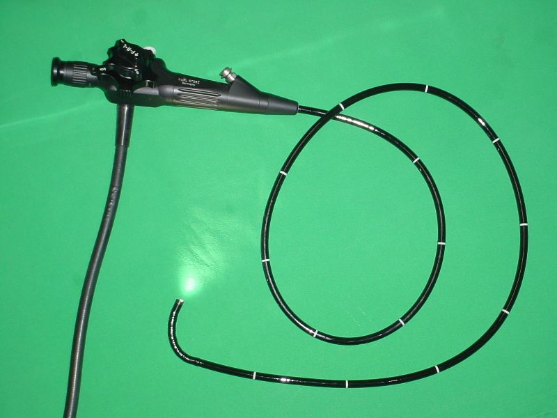
A flexible endoscope. de:Benutzer:Kalumet, CC BY-SA 3.0
The bundle of fibres is in a flexible probe that is inserted into the body, for example at either end of the digestive system, or through blood vessels to view the heart. The tool aperture is used in treatment, for example, to introduce a powerful laser that can be focused on a small area of unhealthy tissue to burn it away.
IMAGING USING RADIOACTIVE TRACERS
Radioactive nuclides emit ionizing radiation. This can be detected by a film, by a Geiger counter or by a scintillation counter. In diagnoses, radioactive nuclides are injected into the bloodstream and their passage through an organ is followed using one of the various detection instruments.
Alternatively, specific areas can be radiolabelled. Biochemical processes involving a series of chemical reactions can be followed using a radioactive isotope of an element in a compound in the series. For example, the uptake of iodine in the thyroid gland is checked by using radioactive iodine.
Here are some of the medical uses for a range of radioactive tracers:
- Elements such as 3H, 24Na, 42K, and 82Br are used as tracers in the general body composition to measure the volume of body fluids and to estimate the quantities of salts (e.g. of sodium, potassium, chlorine).
- Elements such as 32P, 51Cr, 125I, 131I and 132I are used as tracers in the blood to measure the volumes of blood and the different components of blood (plasma, red blood cells) and the volumes of blood in different organs. Also used to locate internal bleeding sites.
- Elements such as 45Ca, 47Ca, , and 99mTc are used as tracers in the bone to investigate absorption of calcium, location of bone disease and how bone metabolizes minerals.
- Elements such as 32P, 60Co, 85Sr, 131I and 99mTc are used as tracers in cancerous tumours to detect, locate and diagnose tumours; 60Co is used to treat tumours.
- Elements such as 99mTc, 131I, 133Xe are used as tracers in the heart and lungs to measure cardiac action: blood flow, volume and circulation. Labelled gases used in investigations of respiratory activity.
- Elements such as 11C, 13N, 15O are used as tracers in the brain: these emit positrons (beta) which interact with electrons to emit gamma rays in positron emission tomography (PET).
The quantity of tracer used must be as small as possible to minimize harmful ionizing radiation. Exposure time is reduced if the substance that is labelled with a tracer is quickly eliminated from the body, or when the isotope has a short half-life.
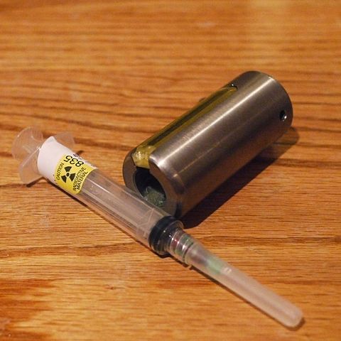
Radioactive Syringe and Shield. Thirteen Of Clubs from Minneapolis, CC BY-SA 2.0
The lifetime of the tracer must be matched to the time scale of any process being studied. Often, a short half-life is useful, as in monitoring blood flow, which can be studied in a short period of time. The tracer should be easy to detect and its position identified accurately.
The best type of emission is gamma (γ) radiation because gamma rays travel easily through matter and cause little ionization. But low-energy beta (β) radiation is also useful. The isotopes listed above are a mixture of beta, positron (β+) and gamma emitters.
Alpha (α) particles are heavily ionizing. This means that alpha emitters are very dangerous and are therefore not useful for producing images.
Technetium-99m
One of the most useful tracers is an isotope of the element technetium:99m43Tc. The ‘m’ indicates that it is a metastable nuclide, meaning that the nucleons in its nucleus are at an energy level higher than in stable technetium. Such nuclei return to normal with a half-life of 6 hours, emitting gamma rays of 140 keV, an energy that makes them particularly easy to detect. The decay produces ordinary technetium-99 which is a naturally occurring radioactive material but has a half-life of 216,000 years, and so it is practically stable, emitting very little radiation.
Radiation detectors in medicine
Most of the basic methods of detecting ionizing radiations have been adapted to medical diagnosis and for monitoring in radiotherapy.
A Geiger counter is used to measure beta radiation, to detect contamination or spillage of radioactive materials. The counter is battery driven and is usually portable. It may include a warning buzzer to show when contamination has reached a dangerous level. Miniature Geiger counters are small enough (20 mm square by 2 mm) to be used inside the body.
An ionization chamber is sometimes used to monitor exposure to radiation and is more accurate than the ordinary film badge. The ionization chamber measures the small current or charge collected when the gas inside the chamber is ionized.
Gamma radiation is of great use in medical diagnosis since it produces very little damaging ionization. For the same reason, it is difficult to detect with a Geiger counter or ionization chamber. The preferred method is to use a series of photomultiplier tubes arranged to form a gamma camera.
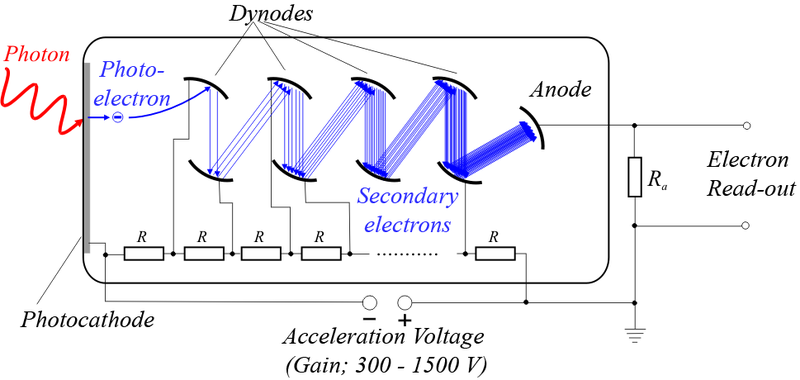
Scheme of a Photomultiplier Tube (PMT). de:Benutzer:Jkrieger, Public Domain
A photomultiplier works in the following way. A gamma ray (emerging through a patient, say) reaches an ionic crystal substance, such as sodium iodide, which emits light when hit by the gamma ray: the crystal scintillates. Because of this effect, the gamma camera scan is called a scintigram. The number of light photons emitted depends upon the gamma ray’s energy. The photons then hit a photocathode, a material that ejects electrons when bombarded by photons in the photoelectric effect. The number of electrons is multiplied using a sequence of charged metal plates called dynodes.
Electrons collide with the plates and have been accelerated enough to liberate secondary electrons from the plates. There may be ten dynodes, each producing four secondary electrons for each incident electron. The electron current is thus multiplied by a factor of 410, that is, by about 106. The whole system is called a photomultiplier tube.
The gamma camera consists of an array of photomultiplier tubes connected to a recording and display system. An image is formed which matches the distribution of gamma emissions from the patient.
POSITRON EMISSION TOMOGRAPHY (PET)
Certain isotopes decay by emitting positrons (positive electrons), the antimatter twin of the electron. In diagnosis, positron-emitting isotopes such as oxygen-15, carbon-11 and nitrogen-13 are used to label compounds that are injected into the body. The compounds chosen are those which tend to collect in specified parts of the body. Once there, the emitted positrons immediately collide with electrons and they annihilate each other, emitting a pair of gamma rays. The patient is surrounded by gamma photon detectors (similar to the gamma camera described above. The output from the gamma detectors is fed to a computer which uses the data to construct ‘real-time’ artificially coloured images on a monitor.
The technique is particularly useful for imaging delicate structures such as that of the brain. For example, when the brain is active the most active parts increase their use of glycogen, as a source of energy. Glycogen labelled with oxygen-15 can then show which parts of the brain are most active when doing different activities such as mathematics, reading, listening to music, etc.
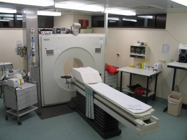
ECAT-Exact-HR--PET-Scanner. Jens Maus, Public Domain.
PHYSICS IN MEDICAL TREATMENT
Many physical diagnostic processes can also be adapted to provide treatment, known as therapy.
THERAPY USING ULTRASOUND
Ultrasound is used to heat small volumes of tissue and destroy small tumours, malignant groups of (cancerous) cells. This requires more intense ultrasound beams than are used in diagnosis, but causes little or no harm to surrounding tissue when a wide beam is accurately focused, or when several beams meet at the point being treated.
Bladder stones can be shattered by the resonance effect when the ultrasound frequency matches their natural frequency of vibration. I have explained in the previous article that cavitation, produced when small air bubbles absorb energy and expand can be harmful. But used carefully, cavitation also promotes wound healing and the repair of damaged bones.
THERAPY USING IONIZING RADIATIONS
Ionizing radiations can kill living cells and both X-rays and radiations from radioactive nuclides are used to treat malignant (cancerous) tumours in radiotherapy.
For treating tumours deep within the body, higher-energy X-ray photons are required. Lower-energy photons are more easily absorbed in soft tissue: they fail to reach deep into the body and may also cause damage to the tissues that absorb them.
High-energy
X-rays are produced by using high voltages, of up to 2 MV compared with the 120
kV used to produce diagnostic X-rays. The output beam of high-voltage tubes
contains a wide range of photon energies and the lower-energy photons are
removed by metal filters (aluminium, tin, lead and gold). The X-rays are delivered
by several beams which converge on the site of the malignant cells. This
reduces the harm to surrounding tissue. Alternatively, a single beam can be
used while the patient is rotated about the target point.
Till next time, I remain my humble self, @emperorhassy.
REFERENCES
https://en.wikipedia.org/wiki/Medical_optical_imaging
https://www.ncbi.nlm.nih.gov/pubmed/9534736
https://www.nibib.nih.gov/science-education/science-topics/optical-imaging
https://www.sciencedirect.com/topics/biochemistry-genetics-and-molecular-biology/radioactive-tracer
https://en.wikipedia.org/wiki/Radioactive_tracer
https://www.youtube.com/watch?v=7mSR--zJGv0
https://en.wikipedia.org/wiki/Technetium-99m
http://videolectures.net/cernacademictraining09_delguerra_urdm/
https://en.wikipedia.org/wiki/Positron_emission_tomography
https://www.radiologyinfo.org/en/info.cfm?pg=pet
https://en.wikipedia.org/wiki/Therapeutic_ultrasound
https://www.physio-pedia.com/Ultrasound_therapy
https://en.wikipedia.org/wiki/Radiation_therapy
https://www.ncbi.nlm.nih.gov/books/NBK232715/
https://opentextbc.ca/physicstestbook2/chapter/therapeutic-uses-of-ionizing-radiation/
This post has been voted on by the SteemSTEM curation team and voting trail. It is elligible for support from @curie and @utopian-io.
If you appreciate the work we are doing, then consider supporting our witness stem.witness. Additional witness support to the curie witness and utopian-io witness would be appreciated as well.
For additional information please join us on the SteemSTEM discord and to get to know the rest of the community!
Thanks for having added @steemstem as a beneficiary to your post. This granted you a stronger support from SteemSTEM.
Thanks for having used the steemstem.io app. You got a stronger support!
Hi @emperorhassy!
Your post was upvoted by Utopian.io in cooperation with @steemstem - supporting knowledge, innovation and technological advancement on the Steem Blockchain.
Contribute to Open Source with utopian.io
Learn how to contribute on our website and join the new open source economy.
Want to chat? Join the Utopian Community on Discord https://discord.gg/h52nFrV
I didn't know about this new advance in the treatment of cancerous tumors, I imagine that it can also be used to destroy benign growths such as fibroids, right?
Posted using Partiko Android
Yes, I guess it can. Thanks for coming by @mike961