Do you know RAMAN spectroscopy? I will give you a brief summary of the techniques used (II part)
My most cordial greetings to the scientific community of steemit, today I come with the continuation of my previous post that was about Raman spectroscopy, at that time I wrote a brief summary about the theory involved in this phenomenon, but this time I will speak on the functioning of the instrumentation used to study Raman spectroscopy and the corresponding techniques used to measure this phenomenon.
As we already know raman spectroscopy provides us with valid information about molecular vibrations within the crystalline structure of a material, this can be used both for the identification and quantification of the samples to be characterized.
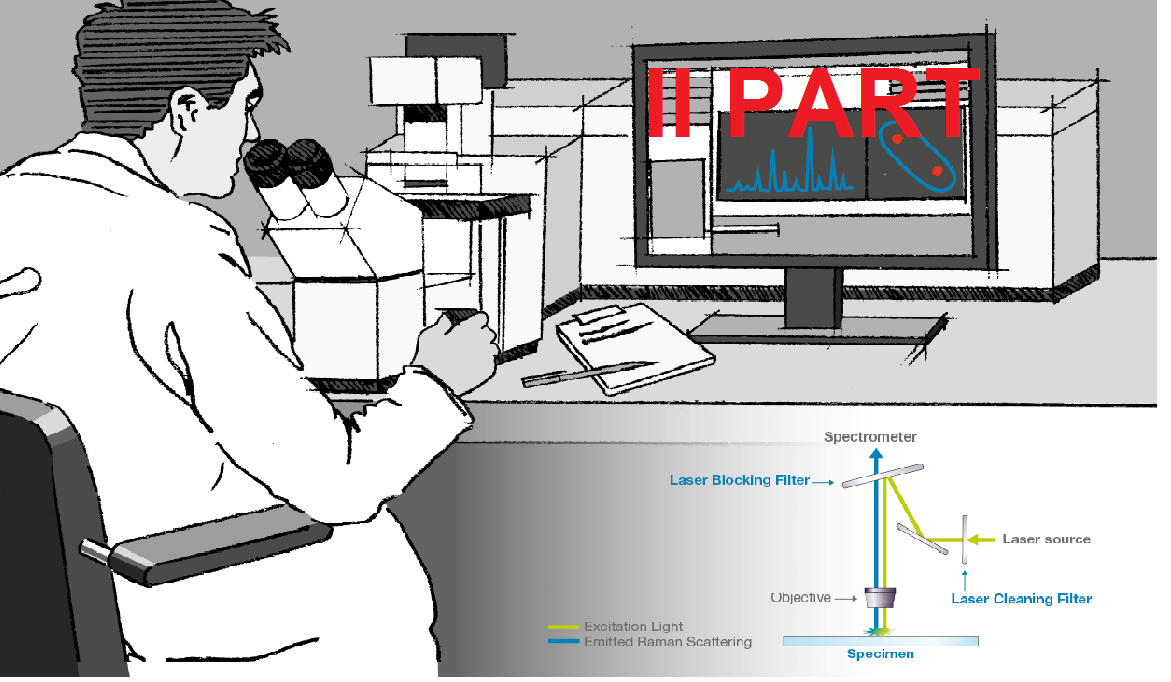
The technique basically consists of illuminating a monochromatic light source in a sample and detecting said scattered light, the scattered light has been the same frequency as the excitation source, that is, most of the scattered light and this is known as the line Rayleigh or elastic dispersion, the small portion of scattered light that remains moves in energy from the frequency of laser light due to electromagnetic interactions or vibrations within the molecule of the characterized material. After this phenomenon occurs and when the intensity of the light shifted as a function of frequency, we obtain the spectrum we are looking for, which is called the Raman spectrum.
Below I will show you the instrumentation needed to perform Raman spectroscopy measurements:
Components of the Raman spectroscopy technique.
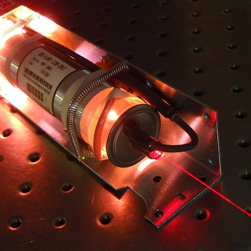
Laser:
It is the source of excitation and is the main component without the light could not disperse, there are many types of lasers but the most recommended to perform this spectroscopy technique is the Helio-Neon red color, which produces a high power, it should be noted that the intensity to produce the Raman scattering is very weak, so a very powerful source of excitation is needed, that is to say that the intensities are greater than the noise of the equipment. The He-Ne laser has a high wavelength of approximately 630 nm, which is why it is the most used source, due to its high degree of phase coherence, which makes it very monochromatic. Other types of lasers used are those of Ar that have a wavelength around 500 nm.
Source
Source
Fiber optics:
Is the means by which laser light travels, to reach the sample and excite it.
During its journey there are two ways, one that is where the laser light is guided to the optical head of the spectrometer, and the other way is the one that collects and carries the laser light to the monochromator. Normally in the experiments when we assemble the equipment a length greater than 10 meters with a diameter of 110 μm is used.
Optical head:
It is composed of different devices among them;
The filter, where the wavelength interference occurs, its function is to let the laser component pass at the frequency that we want. It is composed of the nocht and edge filters, which eliminate all the incorect information during the procedure.
The mirror: allows the bidirectional path between light and excitation, its objective is to reflect the light that goes to the sample to be characterized, and then the information is collected back through the optical fiber.
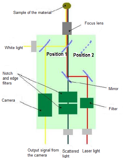
Diagram that includes the components of the optical head.
Lens: focuses the concentration of laser light in the area that the sample is analyzed, it is also responsible for collecting all the scattered light and then perform the corresponding analysis.
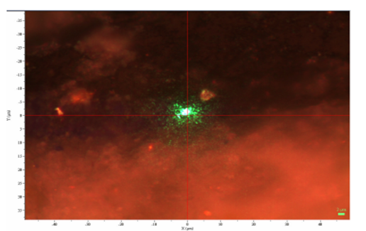
Camera: allows you to control the area where the incidence of the laser occurs correctly, is composed of a peeuqla camera that gives adequate information on the sample image of the material and also has a mirror that can be positioned in the way you want controlled turn by means of movable screw.
Monochromator:
When the laser light is captured by the head of the spectroscope and in turn takes it by means of optical fiber, this signal finally arrives at the monochromator, whose main objective is to capture said signal sent by the laser and separate the components of the specimens. Said component is formed mainly by the grating, inlet and outlet slots, mirrors, collimator.
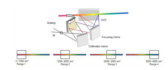
Monochromator shaping scheme.
Source
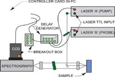
Charge Coupled Device:
That in its acronym better known as the CCD is a capacitor that converts the photons sent by the alser through the optical fiber, after passing through the mirrors in a similar signal, the CCD is responsible for capturing this signal, receives it digitizes the data sent and finally sends it to a computer that shows the light spectra of the material.
Source
Software:
It is the last phase that presents in the measurement system, through this computer program you can visualize the spectral pattern obtained, there is a great variety of programs that help analyze this type of information, the most used are the LabSpec or the SpectraPro , allows to observe and identify bands in the modes vibrations, spectral lines, Fourier transforms, etc.
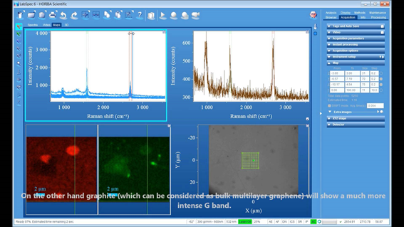
LabSpec computer program.
Source
Some techniques of Raman spectroscopy.
Raman resonance.
Raman confocal microscopy.
Raman Ultraviolet Resonance Spectroscopy (UVRRS).
Improved surface Raman dispersion (SERS) Raman.
Time-Resolved and Pulsed Raman.
To make Raman measurements it is also very important to take into account other additional factors, that is, to perform a good calibration of the equipment, because most of the errors presented in this type of characterization are due to said errors when assembling the equipment, that is why I see myself in the need to explain some tips to be able to obtain spectra desired by us without any type of errors and that at the moment of the analysis, the studies are of total reliability.
Source
The most common error when calibrating the equipment is given by the incorrect movement of the monochromator, since moving it incorrectly becomes the axis of the abscissa, which is the wavelength. The incorrect mobilization produces a horizontal displacement to the measured spectrum, resulting in errors when identifying colors of the measured spectra, since their correct position is that of the bands with respect to the origin.
To avoid this type of inconvenience when interpreting the results is to use known Raman spectra and take them as a pattern such as diamond or ultramarine blue, whose fundamental bands are known and easy to position.
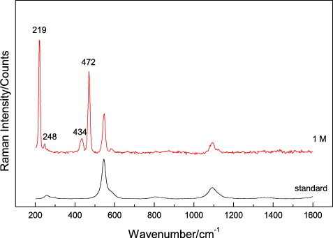
Raman spectrum of ultramarine blue compared with another characteristic spectrum.
Source
But the main calibration method used by most scientists working with spectroscopy, is the use of the Rayleigh component, this is to center this characteristic peak at the origin of the normal wave number.

Calibration through the Rayleigh band.
Advantages of Raman spectroscopy.
This technique has infinite applications such as: food processing, cell treatment, polymer manufacturing, applications in the oil industry, pharmaceutical, among others.
Source
The greatest advantage it has is that no specific treatment is needed for the preparation of the samples to be characterized, since its simplicity allows to inspect with other comparative techniques.
The Micro-raman technique can also analyze the chemical composition of different compounds, either organic or inorganic, and the best thing is that there is no need to destroy the samples for this technique as others, because after characterizing Raman, the Samples can be used in the same way for another type of specific characterization.
Another advantage is that solid, liquid and gaseous compounds can be analyzed.
Other techniques such as X-ray diffraction only allows you to determine the crystalline chemical composition, whereas Raman offers you to characterize any type of material no matter what its state.
In the same way you can study amorphous compounds, organic solids and ordered.
You do not need a vacuum chamber that is expensive and you need a difficult and annoying care, no matter if you are exposed to the environment in the same way you can characterize the spectra, as long as you take the necessary measures of calibration when measuring .
Some applications.
Semiconductors:
It is widely used in the manufacture of synthetic diamond, since it is the only technique that can identify this type of semiconductor compound, it allows to determine the contamination in the contacts of the sample, used in turn in various components of the circuits of electrical appliances.
Biomedical:
It allows to identify different types of cancer: cervix, skin, breast, oral).
Studies in the eye (cornea and fluids).
Alteroclerosis inter-cornaria.
Bone studies, composition, mechanics and their status.
Transdermal drug studies.
Oil industry:
Solvent extraction, aggregation.
BTEX production materials.
Processing of aromatics.
Hydrocarbons.
Gasoline.
And many types of polymers.
Forensic Medicine:
Raman is a very important and extremely powerful tool for the study of forensic medicine, since different and very small samples can be analyzed without the need to destroy them.
Its different applications in this area are: drug abuse, identification of explosives, and chemical substances found in clandestine laboratories including: inks, hair, paints, among others.
Pharmaceutical industry:
Crystallization.
Drying.
Monitoring of new medicines.
Identification of polyform form.
Real-time analysis of chemical reactions.
Application in foreign materials:
Chemical analysis by the composition of the foreign material.
Analysis of organic molecules in the soil.
Identification of mineral phases.
Classification of rocks.
Oxidation state of the elements that are in the ground.
Among others.
Currently, Raman spectroscopy is widely used to identify strange compounds that are not found on planet Earth, it can be used lately for studies of other planets such as Mars, it is a powerful tool for this type of studies and in the future with the progress of the Technology can become the main tool to determine if life exists or not on another planet.
Other apps:
Art and archeology allows the identification of real and false objects.
Similarly in the gemology, verify authentic materials and their origin.
Geology, identification of different minerals.
Determination of different compounds in the environment.
For the alimentary sciences, to be able to corroborate the content of each food.
To conclude we can say that Raman spectroscopy is a very useful tool to be able to identify chemical composition of different compounds or materials, either liquid or solid. It is a very powerful tool to be able to observe small organisms, and the best thing is that you do not need to damage the sample to be able to take your measurements. also has various applications and could be said that in all fields of science, is a very old technique but over the years has been perfected and for scientists is still the main tencica of characterization of materials.
The references with which support was given to the writing of the post are linked to the images.
Other sources of research support:
Thank you very much to the @steemstem team for the support received and the acceptance to your community.

Very interesting read. Great job. I have a background in physics so I managed to understand everything.
You might be interested in a similar series I made on fusion.
I am very happy that the post was of your liking and you understood it, for me it is gratifyingly
Very nice, you got the vote from me for both parts
Thank you very much friend :)
Excelente post. Saludos.
Thanks @rnunez09