THE AUTOMATIC TISSUE PROCESSOR MACHINE
INTRODUCTION
Machines are devices that makes our work easier for us and it does it so efficiently.
• Imagine what the world will be like without machines. Now, machines have found their way into our laboratories, as they help to improve standard health practices.
One of such machine is the –AUTOMATIC TISSUE PROCESSOR. A machine routinely used in the histopathology laboratory.
WHAT IS THE AUTOMATIC TISSUE PROCESSOR MACHINE (ATPM)?
A tissue processor is a device that prepares tissue samples for sectioning and microscopic examination in the diagnostic laboratory.
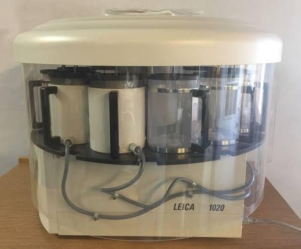
Source
Microscopic analysis of cells and tissues requires the preparation of very thin, high quality sections (slices) mounted on glass slides and appropriately stained to demonstrate normal and abnormal structures.
The ATP machine plays a big role in the preparation of the tissue by passing them through various chemicals; a major process called TISSUE PROCESSING
BRIEF HISTORY OF TISSUE PROCESSOR
Tissue processing has been in existence as far back as in the late 18th century. Major breakthroughs about the basic components of tissue were made possible because of tissue processor.
Disease investigation have also been made easy by subjecting tiny piece (biopsy) of the tissue or organ to some special chemical treatments.
Tissue processing was done manually in the 18th and 19th century. A cruel long process that took days and sleepless nights to achieve this feat. This discomfort forced scientists into looking for a better and a more efficient way to process tissues.
The first automatic tissue processors were introduced during the first half of the 20 th century. In the USA, they were produced under the name of Auto-Technicon and in the UK under the name of Histokine, and later by other companies.
These devices have slowly evolved to be safer to use, handle larger specimen numbers, process more quickly and to produce better quality outcomes.
TISSUE PROCESSING OVERVIEW
The ATPM works by following through an already established processing steps.
Tissues to be processed are cut into small pieces to ensure the tissue fits into the tissue cassettes
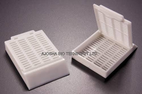
Source
Smaller tissues (2-4 um) will be processed faster than the whole tissue or organ.
These tissue cassettes are packed into the oscillating tissue basket to tissue prior to fixation.
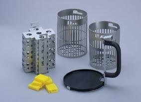
Source
Pic of stainless tissue basket
(i) FIXATION – this is the process of preserving or fixing tissues by passing them through chemicals called fixatives. The fixatives will help protect the tissue from decay and autolysis. Routine fixative of use is 10% formalin
(ii) DEHYDRATION – this is the process of removing water molecules from the tissue by passing the tissue through ascending grades of alcohol. E.g methanol, acetone, 70-100% alcohol
(iii) CLEARING – this is the process of removing alcohol from the tissue by passing it through chemicals that will remove the alcohol molecules. These agents are called clearing agents. Xylene is mostly used for clearing.
(iv) INFILTRATION – this is the process of filling intracellular spaces left in the tissue by paraffin wax. This will help confer a bit of rigidity to the processed tissue.
(v) EMBEDDING- this last step is manually done. This has to do with immersing the processed tissue into a mould containing liquid paraffin wax. This is for external support so that the tissue won’t crumble during microtomy
PARTS OF THE ATPM – using TP 1050 Leica model
(i) Oscillating tissue basket

Source
(i) 10 beakers or jars
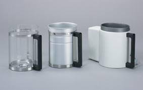
Source
The transparent beakers is for processing fluids
(ii) 2 thermostatically controlled beakers

Source
The coated beaker is for parrafin wax
(iii) An electric rotor at the base
(iv) Lifting mechanism
(v) Time disc and alarm system
(vii) Control unit - with display screen and control buttons
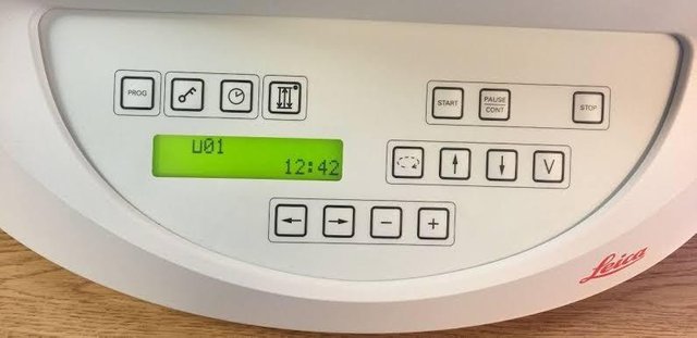
Source
WORKING PRINCIPLE OF AUTOMATIC TISSUE PROCESSOR MACHINE – TP 1050 Leica processor model
Most ATPMs are easy-to-program interface. The Leica processor model has ten 1.8L (60.9oz.) reagent beakers and two 1.8L (60.9oz.) wax baths.
The tissue basket oscillates up and down in each station at three-second intervals to ensure thorough and even mixing of the reagents and optimum tissue infiltration.
Infiltration time is separately programmable for each station. Up to nine programs may be run with immediate or delayed starting times.
When it’s time for tissue to be transferred to the next beaker or jar, the cover of the machine is raised up, and the lifting mechanism carefully removes the tissue basket and gently transfers it to the next beaker.
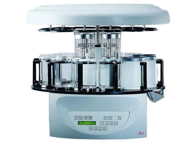
Source
When the infiltration time for any particular station is exceeded, a warning message will display, indicating the station number and excess time. Controls are arranged by functionality with an LCD to indicate operational parameters. Reagent container lids have seals to minimize operator exposure to hazardous fumes.
Tissue basket immediately immerses in a station in the event of power loss to protect samples from drying out. When power is restored, program will resume. In the event of long-term power failure, wax is liquified. Capacity of tissue basket is 80 cassettes.
Vacuum configurations hasten infiltration, allowing pressure to be applied to any station in either manual or automatic operation. Fume control configurations extract fumes with a fan and pass them through an internal carbon filter.
For added efficiency, these models feature a two-part containment shield surrounding the reagent container platform.
ATPM – processing time schedule
Processing schedule varies and it depends oh the following:
(i) Nature and size of tissue
(ii) Urgency
Beaker I – fixative (formalin) 1-2 hours
Beaker II – fixative 1 hour
Beaker III – fixative. 30- 45 minutes
Beaker IV – 70% alcohol. 30 minutes
Beaker V – 90% alcohol. 30 minutes
Beaker VI – Absolute alcohol. 1 hour
Beaker VII – Absolute alcohol. 1 hour
Beaker VIII – Methanol 30 minutes
Beaker IX – Xylene. 1-2 hours
Beaker X – Xylene 45 minutes – 1 hour
Wax bath I (done at 45°c) 2 hours
Wax bath II. 2 hours
Remember that the nature of tissue, size and urgency determines the processing time schedule.
All the processes are performed by the ATPM from start to finish except embedding which is best done manually. This wasn’t possible in the 18th and 19th century.
ADVANTAGES OF ATPM
(i) It’s very efficient
(ii) Saves time and energy to operate
(iii) Cost effective and user friendly
(iv) Can process different tissues same time
(v) The machine does the transfer of tissue from one bath to another. Before now, this wasn’t possible
CONCLUSION
Automatic tissue processor has made tissue processing easy, enjoyable and stressful. I personally enjoy operating this beautiful machine.

SELECTED REFERENCES
- Clayden EC. Practical section cutting and staining. Edinburgh: Churchill Livingstone, 1971.
- Carson FL. Histotechnology . 2nd ed. Chicago: ASCP Press, 2007.
- Hopwood D. Fixation and fixatives. In Bancroft J and Stevens A eds. Theory and practice of histological techniques . New York: Churchill Livingstone, 1996.
- Winsor L. Tissue processing. In Woods A and Ellis R eds.
Laboratory histopathology . New York: Churchill Livingstone, 1994;4.2-1 - 4.2-39.
Fully knowledgeable post.....
Thank You