Epitel Hücrelerimizde Gizlenen Geometri - SCUTOID(Geometry Stored in Our Epithelial Cells - Scutoid)
27 temmuzda Nature Communications'ta yayınlanan bir makale ile bilim insanları yeni bir geometrik şekil keşfettiklerini açıkladılar. Projenin başında Luis Escudero isminde bir biyolog var. Epitel hücrelerinin şeklini inceleyen Escudero, bunları ilk önce prizma olarak düşünmüş ardından frustum olabileceği yönünde çıkarımda bulunmuş. Ancak daha sonra bu şekillerin epitel hücrelerinin şekli olamayacağına ikna olmuş. Çünkü epitel hücreleri hem sıkı hem de esnek bir yapıya sahipler. Prizma ve frustrum bu dinamizme dayanacak şekiller olmadığı için konuyu matematikçilere taşımış Escudero.
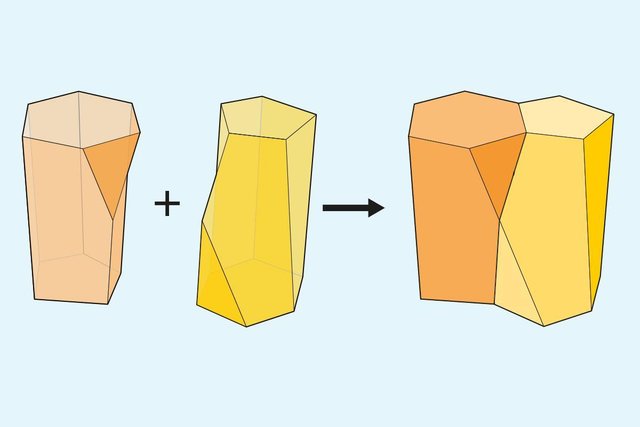
Matematikçiler hücrelerin şekillerinden istifade ederek o şekli ortaya koyan yeni bir formül ile yeni bir geometrik şekil bulduklarını duyurdular: ''SCUTOID''
Çalışmanın raporundan bir grafik
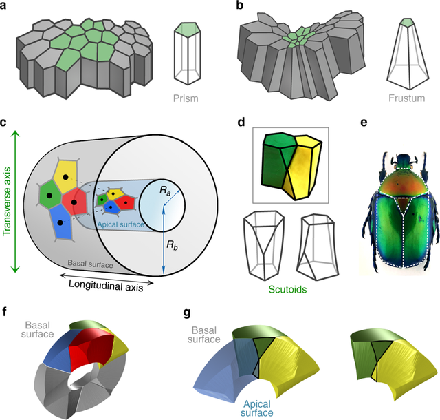
Eğri epitel için bir matematiksel model, yeni bir geometrik katı ortaya çıkarır. a Düzlemsel kolumnar / kübik tek tabakalı epitelyumu temsil eden şema. Hücreler prizmalar olarak basitleştirilmiştir. b Bir kolonar / kübik tek tabakalı epitelde bir invaginasyon veya katlamayı gösteren şema. Hücreler, frusta olarak basitleştirilecek "şişe şekli" olarak adlandırılır. c Bir epitel tüpü için matematiksel model. Bir silindirin yüzeyinde (epitel tüpünün apikal yüzeyini temsil eden) bir Voronoi diyagramı çizilir. Her bir Voronoi hücresinin tohumları bir dış silindirde (epitelyum tüpünün taban yüzeyini temsil ederek) yansıtılır. Bu bir topolojik değişimi, bir hücre interkalasyonunu indükleyebilir. Sarı ve mavi hücreler, apikal yüzeyde değil, bazal yüzeyde komşudur. Karşılıklı kırmızı ve yeşil hücreler için oluşur. Ra, silindirlerin merkezinden apikal yüzeye kadar yarıçap. Rb, silindirlerin merkezinden taban yüzeyine kadar yarıçap. d Bir geçişe katılan iki scutoid ve scutoid katıları için iki şemaları gösteren kil şekillerinin modellenmesi. Scutoidler, iki tabana farklı bir düzlemde en az bir tepe noktası bulunması ve eğri yüzeylerin mevcut olması ile karakterize edilir. e Cetoniidae ailesinden bir Protaetia speciose türünden bir meyve böceğinin dorsal görünümü. Beyaz çizgiler, scutum, scutellum ve kanatların benzerliklerini scutoidlerin şekliyle vurgulamaktadır. Nicolas Gompel'den izin alarak konulmuştur. f Rb ∕ Ra = 2.5 olan bir tüp oluşturan hücrelerin 3D rekonstrüksiyonu. Dört hücreli motif (yeşil, sarı, mavi ve kırmızı hücreler) bir apiko-bazal hücre interkalasyonunu gösterir. g Mavi ve sarı hücrelerin apikal kısımda nasıl temasa geçtiğini gösteren, ancak bazal kısımda olmayan apikor-bazal geçiş detayı. Şekil ayrıca scutoidlerin içbükey yüzeyler sunduğunu da göstermektedir. Git
Bir video:
Bir bağlantı: Nature
For English:
On July 27, an article published in Nature Communications explains that scientists are discovering a new geometric shape. At the beginning of the project is a biologist called Luis Escudero. Escudero, studying the shape of epithelial cells, made a conclusion that he might first think of them as a prism and then a frustum. However, later on these forms were convinced that the epithelial cells could not be shaped. Because epithelial cells have both a tight and flexible structure. The prism and the frustum weren't the shape to endure this dynamism and Escudero were carried this problem to the mathematicians.
Mathematicians announced that they had found a new geometric shape with a new formula that revealed that shape by taking advantage of the shapes of the cells: ''SCUTOID''
A graph from the report of the work
A mathematical model for curved epithelia uncovers a novel geometrical solid. a Scheme representing planar columnar/cubic monolayer epithelia. Cells are simplified as prisms. b Scheme illustrating an invagination or fold in a columnar/cubic monolayer epithelium. Cells adopt the called “bottle shape” that would be simplified as frusta. c Mathematical model for an epithelial tube. A Voronoi diagram is drawn on the surface of a cylinder (representing the apical surface of the epithelial tube). The seeds of each Voronoi cells are projected in an outer cylinder (representing the basal surface of the epithelial tube). This can induce a topological change, a cell intercalation. Yellow and blue cells are neighbours in the apical surface but not in the basal surface. The reciprocal occurs for red and green cells. Ra, radius from the centre of the cylinders to the apical surface. Rb, radius from the centre of the cylinders to the basal surface. d Modelling clay figures illustrating two scutoids participating in a transition and two schemes for scutoids solids. Scutoids are characterized by having at least a vertex in a different plane to the two bases and present curved surfaces. e A dorsal view of a Protaetia speciose beetle of the Cetoniidae family. The white lines highlight the resemblance of its scutum, scutellum and wings with the shape of the scutoids. Illustration from Dr. Nicolas Gompel, with permission. f 3D reconstruction of the cells forming a tube with Rb∕Ra=2.5. The four-cell motif (green, yellow, blue, and red cells) shows an apico-basal cell intercalation. g Detail of the apico-basal transition, showing how the blue and yellow cells contact in the apical part, but not in the basal part. The figure also shows that scutoids present concave surfaces. Go
A video:
A link: Nature
İleri okumalar için (For further reading):