Life Explorers - The Human Senses Part III: Touch

Many things around us and with us happen we take for granted. We see, feel, taste, hear and smell unknowingly how it works. As we were children we called anything into question and now accept reality as it is. We lost the magic in all these wonderful things. This series addresses the wonder of natures and will bring its magic back to light. The third part of the Life Explorers - The Human Senses deals with the sense of touch. We feel the wind touching our skin, we feel pain when we cut our finger, when someone caresses your cheek you feel it. But what actually is feeling?
To be able to understand how the sense of touch works we will investigate the somatosensory system of the human body, which is able to detect physical stimuli.
The largest organ – The skin
In order to understand how the different perceptions like temperature, pressure and pain operate, we will have a look at the largest organ of the human body, the skin. The skin contain different receptors which are essential for surviving, making the skin one of the most important organ. We will further discuss the different receptors the skin contains. The following figure shows a cross section of the human skin with it's receptors:
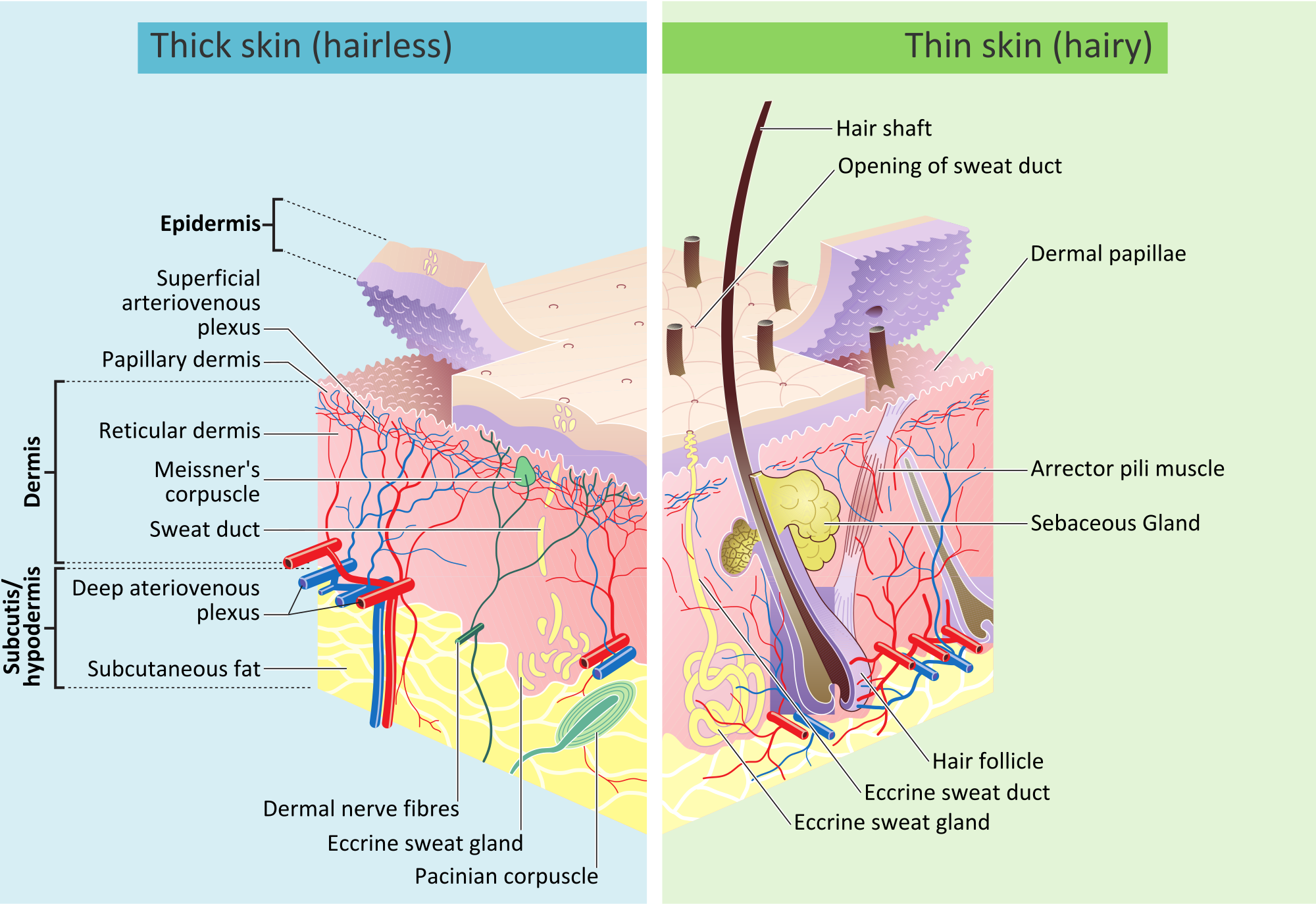
The human skin has three layers. The epidermis, dermis and the subcutis. Most sources speak of the first two layers but do not consider the Subcutis as a skin layer. We will only investigate the first two layers, epidermis and dermis, since these two play the main role in the sensation.
Epidermis:
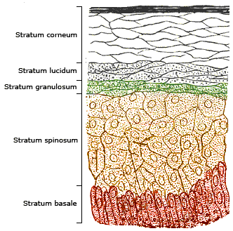
The epidermis is composed of five different layers. The corneum cell layer, lucidum cell layer, granulosum cell layer, spinosum cell layer and the basale cell layer. Since the epidermis is the top layer of our skin and has contact to the environment is has to have specific properties to protect the skin from possible harm. The deepest layer of the Epidermis is the basale cell layer, containing three important types of cells: The keratinocytes stem cells, the melanocytes and the Merkel cells.
To get a better glimpse of how of skin works I will explain these cells in short. The keratinocytes stem cells are producing keratinocytes by cleavage of cells. This cell division causes new cells to move to the skin surface. By doing so the keratinocytes change their form. This happens because the further the keratinocytes move the less they can the blood supply these cells. They start to die and move further up. Reaching the top we can observe them as cornea, also known as horny skin.
The melanocytes are responsible for our skin color by producing melanin, a color pigment. There are two different types of melanin responsible for our skin color, eumelanin and pheomelanin. Eumelanin causes a brown to black color whereas pheomelanin is causing a yellow to red hue. The darker your hair is the more eumelanin you have, for instance. Besides the skin color melanin protects us against sunlight, by absorbing 99.9% of UV radiation. This absorption is protecting the deeper laying skin layers from radiation. At the same time sunlight is stimulating the production of more melanin, this is also commonly known as tanning. Another cell we will have a closer look at are the Merkel cells. These are important for our sensation since they are receptor cells, but we will come to the receptors later.
We classify the following receptor types:
| Mechanoreceptors | Merkel, Meißner, Pacini | Touch, Vibration |
| Thermoreceptors | Krausesche, Ruffini | Hot/Cold |
| Nociceptors | Free nerve ends | Pain, extreme temperture (+45 C) |

Dermis
Before we further investigate these receptor cells we will take a look at the dermis, the second layer of our skin. The dermis is under the epidermis and contains most of the receptors, blood and lymph vessels as well as cutaneous glands and hair cells. The cutaneous glands are divided into sebaceous and sweat glands, providing our skin with sebum (tallow) and sweat, which cools down our body. Consisting of conjunctive tissue like collagen and elastin, the dermis is responsible for the flexibility and strength of our skin. The older we become, the more winkles we have. This is due to the degeneration of elastin and collagen as well as the decline of subcutaneous fat. Because the epidermis has no blood vessels the dermis is providing the top layer with nutrient. The dermis itself is separated into two layers, the papillary and the reticular layer. The papillary layer connects the epidermis with the dermis and gives our top skin a certain pattern. The papillary connection is the reason we have unique finger prints.
The epidermis has an area of 2 qm and together with dermis and subcutis together make 11-15 Kg of our body weight.

The receptors – Biochemistry that shows how complex the human body works
Now that we know the general structure of the human skin, we will introduce the scientific part, or as I like to say: The magic. In our previous Life Explorer article we stopped at this point in order to prevent information overload. In order to understand how we feel with our body we need to understand the basic fundamentals of nerve transmissions. We will investigate how pain is created and see how pain is nothing more than a chemical reaction.
Nociceptors – Our friends that hurt us
How does pain occur? We can cut our finger for instance, fall badly or have inflammation. There are many reason why, but they all have the same thing in common. They are based on stimulation of sensory nerves. The moment the knife cuts through our skin we feel pain and reflexively shrug. These are two things we have to distinguish here. First, pain is a conscious sense, whereas the reflex to pull back our finger is a vegetative reflex and therefore unconscious.
Pain is cause by the nociceptors in our dermis. Nociceptors are free nerve endings outbound from the spinal cord that transform stimuli into electrical signals to the brain. We are able to feel different types of pain: somatic and visceral pain. Somatic pain is considers as external influence that harm our tissue whereas visceral pain comes from the inside of our body. There are two different types of these receptors: C-fiber and Aδ. The main difference between these two types of fibers are the speed of signal transmission. The C-fibers conduct a signal with 1 m/s whereby the Aδ nociceptors can conduct with a speed up to 30 m/s. The different speed causes different pain. The first pain we feel is caused by the Aδ and is followed by a deep pain, caused by the C-fibers.
The following figure shows respectively the way a signal makes from the receptor ending through the spinal cord which ends in certain areas of the cortex.

Nociceptors are usually on standby, luckily, otherwise we would feel continuously pain. In order to send a signal to the brain, nociceptors have to be activated. Nociceptors are sensitive to three kinds of noxic stimuli: mechanical, chemical and thermal. We will have a look into mechanical stimuli. To understand how a neuronal signal is created we need to know what a neuron is and what causes the stimulation.
The following illustration shows the structure of a neuron:

The main parts a neuron consists of are the dendrites, the axon and the axon terminal. For our further understanding of transmitting a signal we will only have a look on the dendrites and the synapses. Synapses build the connections between two neurons. They are responsible for transferring neuronal stimuli to the next neuron. Using the example of cutting the finger, the synapses would transfer the pain from the finger to the brain.
Stimulus-Response Pathway
Stimuli are electrical signals that are transfers through neurons with the help of synapses. We will introduce the following terms: resting and action potentials.

Resting potential:
Every cell membrane logically has two sides which are separating two compartment. These two sides have a different concentration of molecules, here ions, causing a potential difference. The Sodium/Potassium-Pump in the membrane is bringing 2 K+ Ions into the cell and 3 Na+ ions outside of the cell. In sum one positive charge is leaving the cell. This creatives a negative membrane potential of -60 to -70 mV.
In order to transfer a signal we need to bring this cell into an action potential. This happens when a neuronal signal approaches our cell and changes the potential. When it reaches -55 mV the cell is opening its Na+ gateways and sodium ions move into the cell, changing the potential to +40 mV. The cell got depolarized. This action potential causes Ca++ channels to open which then releases neurotransmitters. These Neurotransmitter however cause another action potential in the next neuron.
As mentioned we have three different stimuli for the nociceptors: mechanical, chemical and thermal. When a rough force acts upon our skin or tissue, this mechanical force can open the Na+ channels, causing an action potential. The longer a nociceptor is active the more sensitive the area around it. The threshold to release a potential is lowered and the area around our injury is more sensitive. This is the reason why inflammations hurt when we touch the skin.
Author's Note:
The Life Explorer Series is a community magazine that brings together writers to post about a variety of topics. All topics and authors using the lifeexplorer tag or title are part of this group and have permission to post under the heading Life Explorer. If you would like to write with the Life Explorer series about a topic, reach out and get in chat contact with @timsaid to learn more.
Make sure to catch all these Life Explorer authors:
@prufarchy
@yogi.artist
@disofdis
And if you've missed the previous edition, check out the first Life Explorer post:
Life Explorers - The Human Senses Part I: Sight
Life Explorers - The Human Senses Part II: Hearing

Thanks, please keep it coming.
Surprised, the human organism works.
Good read. Thank you!
very interesting! Upvoted!
Facinating, thanks for the read man!
Thank you for the kind words! :) Feedback always helps to improve
a good details of explanation
Hoy he aprendido cosas interesante con su post, jamas me lo hubiera imaginado que el órgano mas grande era la piel. Muy buen trabajo, lo que ud ha realizado. Muchas Gracias
Cool article !!!
I wonder, why majority of current experiments in designing haptic interfaces concentrate around mechanical vibrations at certain frequencies vs. electric stimulation, which would make me think have better control over neurotransmitters' reactions.
interesting read, thank you:)