New Microfluidic Chip Will be Able to Replace Muscle-Nerve Connections
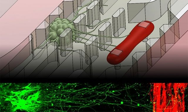
Massachusetts Institute of Technology engineers have created a microfluidic device that is able to precisely imitate the neuromuscular junction, which is a crucial connection where nerves and muscles come together as one. The device is the size of a quarter and is made up of a single muscle strip and a tiny set of motor neurons. Researchers are able to influence the interactions, viewing them within a realistic and three dimensional matrix.
Researchers genetically modified neurons within the device in order to respond to light. They were able to precisely stimulate cells by shining light directly on the inside neurons which lead to signals being sent that excite the muscle fiber. Researchers measured the force exerted by the muscles within the device through a series of twitches and contracts that the device responds to.
The complete results were published in Science Advances and this may help scientists to better understand and identify drugs in order to treat amyotrophic lateral sclerosis (called ALS), which is most commonly called Lou Gehrig's disease, and a long list of other neuromuscular-related conditions.
Sebastien Uzel, leader of the work and graduate student in MIT's Department of Mechanical Engineering says the neuromuscular junction is involved in a lot of very incapacitating and sometimes even brutal and fatal disorders which much has yet to be discovered. His hope is that being able to form neuromuscular junctions in vitro will help us understand how certain diseases function.
Ever since the 1970s, researchers have come up with a number of ways to simulate the neuromuscular junction within a lab. Most of their experiments involved growing muscle and nerve cells in Petri dishes. Unfortunately these environments are not very similar to the conditions of the human body, where muscles and neurons live in complex and three dimensional environments with long distances often separating them
Uzel, postdoc at the Wyss Institute at Harvard University says to think of a giraffe. There are neurons that live in the spinal cord that send axons across very long distances in order to connect the muscles within the leg. In order to build more realistic in vitro neuromuscular junctions, Uzel and his colleagues fabricated a microfluidic device that was made up of two very crucial features: a three dimensional environment and compartments that separated muscles from nerves in order to copy the natural separation that takes place in the human body. The researchers then suspended muscle and neuron cells inside of the millimeter-sized compartments which were filled with gel in order to act similarly to a three dimensional environment.
In order to grow a muscle fiber the team reached for used muscle precursor cells from mice which they differentiated into muscle cells. The cells were injected into the microfluidic compartment where they grew and fused to create a single muscle strip. Motor neurons were also differentiated from a cluster of stem cells. The resulting aggregate of neural cells were put into the second compartment. Before both cell types were differentiated, researchers genetically modified the neural cells so they would respond to light via a technique called optogenetics.
Kamm says light, as opposed to electrodes, offers a pinpoint control of what cells you are able to activate. In the very confined space, electrodes hold the potential of accidentally stimulating cells other than the ones being targeted.
Researchers added force sensing to the device during the final stage of testing. In order to measure muscle contraction, they had to fabricate two small, flexible pillars inside the muscle cells' compartment. The growing muscle fiber could then wrap around this area. When the muscle contracted, the pillar squeezed together which created a displacement that researchers were able to measure and convert to mechanical force.
During experiments done to test the device, Uzel and his colleagues watched as neurons extended axons toward the muscle fiber within the three dimensional region. Once they discovered that an axon had made a connection, they stimulated the neuron with a tiny burst of blue light and watched as the muscle instantly contracted.
Kamm says when you flash a light, you get a twitch. Based on these experiments, Kamm explains that the microfluidic device may be able to be served as a testing ground for drugs that treat neuromuscular disorders and could even be custom tailored to patients on an individual basis. He believes that you could potentially take pluripotent cells from an ALS patient, differentiate them into muscle and nerve cells and then make the whole system for that particular patient. Then you would be able to replicate it as many times as you want, and try different drugs or combinations of therapies to see which is most effective in improving the connection between nerves and muscles.
The device may also be useful in modeling exercise protocols. For example, by stimulating muscle fibers at varying frequencies, scientists can study how repeated stress affects the performance of muscles. Kamm says now with all these new microfluidic approaches people are developing, you can start to model more complex systems with neurons and muscles. The neuromuscular junction is another unit people can now incorporate into those testing modalities.

Like what you read (without annoying ads :) ) ? Then please upvote our posts and follow us for the best daily science news on SteemIt!
Sign me up
Unfortunately it isn't ready for mass production but in a few years this could be a reality for everyone...