A Nitric Oxide Nanosensor
Electrochemical Nanosensor for Real-Time Direct Imaging of Nitric Oxide in Living Brain
Analytical Chemistry, 2011, 83 (21), pp 8314-8319 dx.doi.org10.1021/ac202225n
This paper describes a technique for real-time nitric oxide (NO) imaging in vivo. The promise of easy analysis of this signalling molecule in a living brain could prove beneficial to those studying NO’s biological roles. Nitric oxide gas, which provides important functions as a vertebrate biological messenger has recently been found to play a much greater role in neural function than previously thought. As one of its many functions, NO regulates neurovascular coupling and acts as a neurotransmitter in synaptic plasticity and signaling regulation. However, excess NO in brain tissue is toxic and plays a role in apoptosis and ischemia. NO is releases excessively in many neurodegenerative disorders, such as Alzheimer’ and Parkinson’s disease. Hence, a simple characterization method could be invaluable. In this paper, the authors couple a NO nanosensor with scanning electrochemical microscopy (SECM). This technique is unique in its capability of providing three-dimensional information of NO release in an intact brain, in real-time. The method can show NO release sites, infrared intensity, and relationships between measurements. It can also tell the size and location of related cells.
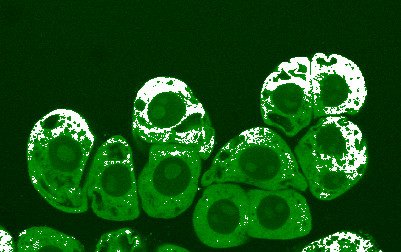
Production and diffusion of NO in cytoplasm, Don J Durzan. Wikimedia Commons
_(white)_in_the_cytoplasm_(green)_of_clusters_of_conifer_cells_one_hour_after_mechanical_agitation.jpg)
Most NO analyses are performed with spectroscopic techniques, but these commonly have limitations with regard to in vivo analysis. Some have low specificity or poor detection limits, while others are disadvantaged with time-consuming steps or toxicity of co-reactants. The paper’s authors found a solution to these issues by developing a Platinum nanopore electrode which is simple and quick to use for in vivo analysis.
The reliability of this NO imaging technique was confirmed with immunohistochemical analysis of a living brain. The NO imaging results were well matched with the corresponding neuronal NO synthase immunoreactive cells. With the SECM technique, a microelectrode measures the electrochemical current as a function of the electrode location while scanning across the sample surface. Local NO concentrations were determined using a planar-type NO nanosensor as the probe tip in SECM. This nanosensor is a glass nanopore Pt electrode created using chemical etching, and electrodeposition to produce a hydrophobic pore environment with high selectivity for NO over interfering species. Pore radii were determined to be less than 500 nm with a scanning electron microscope.
Listen to this: my buddies over there said that I wouldn't be able to start a conversation with the most beautiful boy/girl in the bar. Wanna buy some drinks with some of their money?
To listen to the audio version of this article click on the play image.

Brought to you by @tts. If you find it useful please consider upvoting this reply.
interesting great info have a great day
Is the equipment used to perform this kind of photography expensive? It's pretty neat!
I love reading this article, I learned a lot about how you analyzed our brain, I think you might know more if you are involved with it.@pinkspectre am I right 👉 sir???