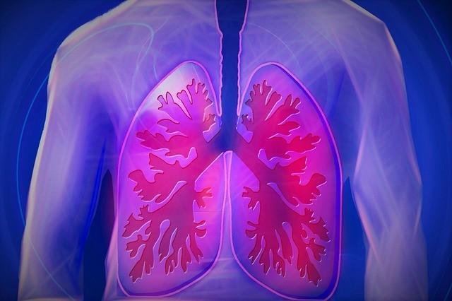
Effects of exercise on the cardiopulmonary ventilation system
Ventilation has any definition but depend on physiology the ventilation is the movement of air between the environment and the lungs via inhalation and exhalation.
Control of Ventilation
The precise mechanism responsible for increased alveolar ventilation during exercise is not well understood. Exercise causes the body to consume a large amount of oxygen and, simultaneously, to produce a large amount of carbon dioxide. Alveolar ventilation increases so much, however, that the concentration of these gases in the body does not change significantly. In addition, no oxygen or carbon dioxide chemoreceptors have been identified on the venous side of circulation, or in the lungs, that could account for the increased alveolar ventilation during exercise. Thus, it is unlikely that the increased ventilation seen in exercise is caused by either of these gases.The control of ventilation refers to the physiological mechanisms involved in the control of breathing, which is the movement of air into and out of the lungs. Ventilation facilitates respiration. Respiration refers to the utilization of oxygen and production of carbon dioxide by the body as a whole, or by individual cells in cellular respiration. Source
It has been suggested that the increased ventilation is caused by neural im- pulses sent to the medulla by way of the following pathways :
- The cerebral cortex sending signals to the exercising muscles may also send collateral signals to the medulla oblongata to increase the rate and depth of breathing.
- Proprioceptors in the moving muscles, tendons, and joints transmit sensory signals via the spinal cord to the respiratory centers of the medulla.
- The increase in body temperature during exercise also may contribute to increased ventilation.
Alveolar Vertilation
During normal quiet breathing, an adult exchanges about 6 L of air per minute. During strenuous exercise, this can increase to 120 L/min, a 20-fold increase. De- pending on the intensity and duration of the exercise, alveolar ventilation must in- crease to (1) supply sufficient oxygen to the blood, and (2) eliminate the excess carbon dioxide produced by the skeletal muscles. The increased alveolar ventila- tion is produced mainly by an increased depth of ventilation (increased tidal vol- ume), rather than by an increased rate of ventilation. During very heavy exercise, however, both an increased depth and frequency of ventilation are seen. The tidal volume is usually about 50 percent of the vital capacity, and the respiratory rate is usually between 40 and 50 breaths/min.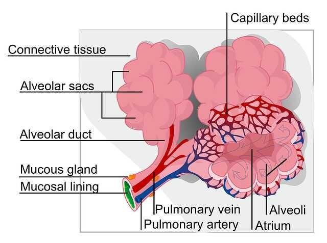
There are three distinct consecutive breathing patterns seen during mild and moderate exercise. The first stage is characterized by an increase in alveolar ventilation, within seconds after the onset of exercise. The second stage is typified by a slow, gradual further increase in alveolar ventilation developing during approximately the first 3 minutes of exercise. Alveolar ventilation during this period increases almost linearly with the amount of work performed. During the third stage, alveolar ventilation stabilizes. When an individual stops exercising, alveolar ventilation decreases abruptly.
During very heavy exercise, the steady-state third stage may not be seen. In fact, when approximately 60 to 70 percent of the maximal exercise level is reached during the linear second stage, alveolar ventilation increases proportionately more than the oxygen uptake. The additional stimulation is thought to be caused primarily by the accumulation of lactic acids in the blood after the anaerobic threshold has been reached. It is suggested that the H+ ions generated by the lactic acids stimulate the carotid chemoreceptors, which in turn send neural impulses to the medulla oblongata to increase alveolar ventilation.
The maximum alveolar ventilation generated during heavy exercise under normal conditions is only about 50 to 65 percent of the maximum voluntary ventilation (also called maximum breathing capacity). This provides the athlete with an important reserve of alveolar ventilation, which may be required in such conditions as short bursts of increased exercise, exercise at high altitudes, or exercise during very hot and humid conditions. Because there is normally a large alveolar ventilatory reserve during exercise, it is not the limiting factor in the delivery of oxygen to the muscles during maximal muscular aerobic metabolism.
Oxygen Consumtion
At rest, normal oxygen consumption is about 250 mL/min. The skeletal muscles account for approximately 35 to 40 percent of the total VO2. During exercise, the skeletal muscles may account for more than 95 percent of the VO2. During heavy exercise, the V02of an untrained person may be more than 3500 mL of O2/mm. The VO2of an elite athlete while running a marathon may be over 5000 mL O2/min.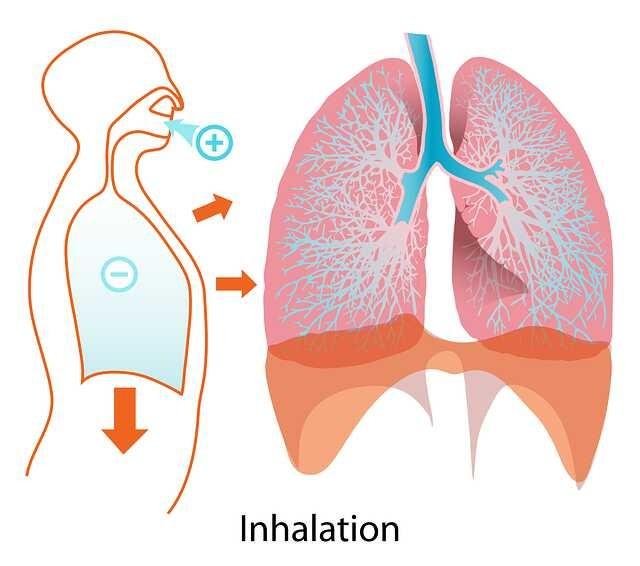
Arterial Blood Gas Levels During Exercise
No significant Pa02, PaCO2 or pH changes are seen between rest and approximately 60 to 70 percent of maximal VO2. During very heavy exercise, however, when lactic acidosis is present, both the pH and PaCO2decline. Although controversy exists, it is believed that arterial acidosis stimulates the carotid chemoreceptors, causing increased alveolar ventilation and promoting respiratory acid base compensation. The PaCO2remains constant during mild, moderate, and heavy exercise.
Oxygen Diffusion Capacity
The oxygen diffusion capacity increases linearly in response to the increased oxygen consumption (V02), during exercise. The oxygen diffusion capacity may increase as much as three fold during maximum exercise. It has been shown that the increased oxygen diffusion capacity results from the increased cardiac output during exercise. The increased cardiac output causes the intravascular pressure in the pulmonary artery and left atrium to increase, which in turn serves to (1) distend the pulmonary capillaries that are not fully dilated and (2) open, or recruit, closed pulmonary capillaries. As more blood flows through the lungs, more alveolar-capillary units become available for gas exchange. This provides a greater surface area through which oxygen can diffuse into the pulmonary capillary blood.
Very comprehensive collection of relevant information! I learn every day, thanks for that!!
You are welcome brother...thank for comment