Did You Know Visualizing The Interior Of The Human Body
The human body is the entire structure of a human being. It is composed of many different types of cells that together create tissues and subsequently organ systems. They ensure homeostasis and the viability of the human body.
It comprises a head, neck, trunk (which includes the thorax and abdomen), arms and hands, legs and feet.
The study of the human body involves anatomy, physiology, histology and embryology. The body varies anatomically in known ways. Physiology focuses on the systems and organs of the human body and their functions. Many systems and mechanisms interact in order to maintain homeostasis, with safe levels of substances such as sugar and oxygen in the blood.
Medical imaging
Medical imaging is the technique and process of creating visual representations of the interior of a body for clinical analysis and medical intervention, as well as visual representation of the function of some organs or tissues (physiology).
Medical imaging seeks to reveal internal structures hidden by the skin and bones, as well as to diagnose and treat disease. Medical imaging also establishes a database of normal anatomy and physiology to make it possible to identify abnormalities. Although imaging of removed organs and tissues can be performed for medical reasons, such procedures are usually considered part of pathology instead of medical imaging.
Visualizing Internal Organs
Modern technological advances have enabled the human body to be visualized and investigated in a wide variety of ways. Imaging creates pictures of internal organs and structures, which may be displayed as digital images on a monitor or recorded on photographic film. Viewing techniques use specialized devices, including endoscopes, to look directly at internal structures of the body.
In the past, the need for exploratory surgery brought with it hospitalization, risk of infection, and discomfort and pain for the patient. Today, however, several technologies and the extensive use of computers permit us to see the interior of the body without surgery.
Here I will give you examples imaging technique of tools commonly used to visual the internal organs, although there are still many other tools that are used. For more Visualizing Internal Organs you can read here.
Computed tomography (CT)
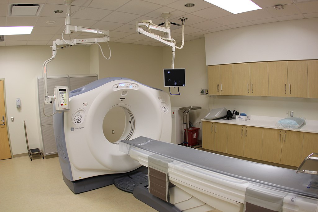
Computed tomography (CT) scanning uses a narrowly focused x-ray beam that circles rapidly around the body. A detector then measures how much radiation passes through different tissues, and a computer constructs an image of a thin slice through the body. Several images may be made at different levels each takes only a few seconds to provide a more complete picture of an organ or part of the body. The images are much more detailed than are those produced by conventional x-rays.
A CT scan, also known as computed tomography scan, makes use of computer-processed combinations of many X-ray measurements taken from different angles to produce cross-sectional (tomographic) images (virtual "slices") of specific areas of a scanned object, allowing the user to see inside the object without cutting. Other terms include computed axial tomography (CAT scan) and computer aided tomography.
Magnetic resonance imaging (MRI)
Magnetic resonance imaging (MRI) is a medical imaging technique used in radiology to form pictures of the anatomy and the physiological processes of the body in both health and disease. MRI scanners use strong magnetic fields, radio waves, and field gradients to generate images of the organs in the body. MRI does not involve x-rays, which distinguishes it from computed tomography (CT or CAT).
MRI has another diagnostic tool that is especially useful for visualizing soft tissues, including the brain and spinal cord. Recent refinements have produced imagesof individual nerve bundles, which had not been possible using any other technique.
The patient is placed inside a strong magnetic field, and the tissues are pulsed with radio waves. Because each tissue has different proportions of various atoms, which resonate or respond differently, each tissue emits a characteristic signal. A com- puter then translates these signals into an image; the entire procedure takes 30 to 45 minutes.
Positron emission tomography (PET)
Positron emission tomography (PET) scanning creates images that depict the rates of physio- logical processes such as blood flow, oxygen usage, or glucose metabolism. The comparative rates are depicted by colors: Red represents the highest rate, followed by yellow, then green, and finally blue representing the lowest rate.
Positron-emission tomography (PET)has a nuclear medicine functional imaging technique that is used to observe metabolic processes in the body. The system detects pairs of gamma rays emitted indirectly by a positron-emitting radionuclide (tracer), which is introduced into the body on a biologically active molecule.
Three-dimensional images of tracer concentration within the body are then constructed by computer analysis. In modern PET-CT scanners, three-dimensional imaging is often accomplished with the aid of a CT X-ray scan performed on the patient during the same session, in the same machine.
Conclusion
From above article we can conclude that these technologies is their cost; they are expensive. However, the benefits to patients are great: Highly detailed images of the body are obtained without the risks of surgery and with virtually no discomfort in the procedures themselves.