How the eye captures images
The eye is a visual instrument in its own right and perhaps more perfect than any instrument that men have ever devised. The complete visual mechanism involves two steps:
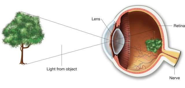
(i) The eye has a lens which being convex in nature produces a real and inverted image of an object on a light-sensitive membrane, called retina, at the back of the eye-ball;
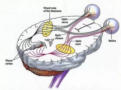
(ii) The sensation of the image is then sent through optic nerves to the brain, which read what the image means.

Structure of the human eye
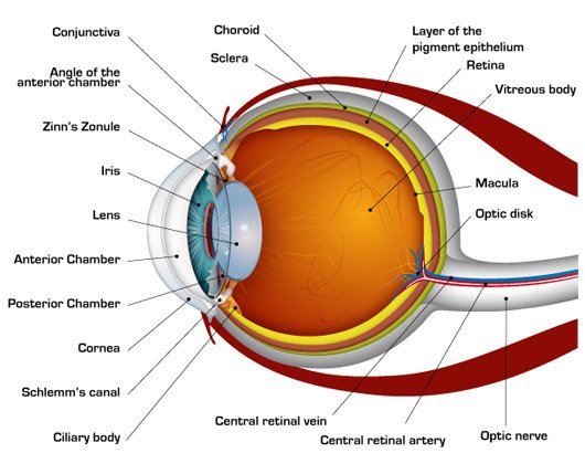
The shape of the human eye is almost spherical. It is placed in bony socket in skull and can be circulated within this socket extensively with the help of the complex action of six muscles. The eye has a white unclear coating made of tough fibrous tissue called the sclerotic. This is the ‘white’ of the eye. The front division of the sclerotic, called the cornea, is transparent with bigger curvature. Light gets in the eye through the cornea. The next layer inside the sclerotic is called the choroid. It is a dark thin membrane made of many blood vessels. The innermost layer of the eye is called the retina where the images are formed. It is also a light sensitive membrane which directly has connection with the optic nerve. The presence of black pigments in the choroid prevents lights from being reflected back on to the retina again.

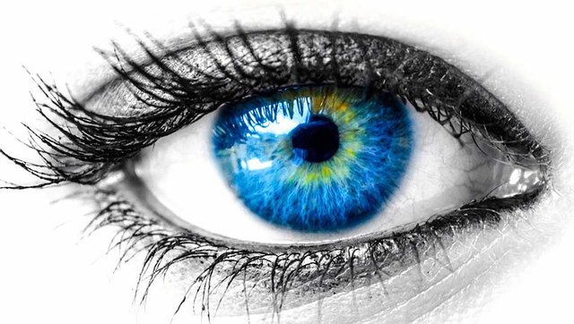
A narrow slit like anterior chamber, which contains aqueous humor, is found behind the cornea. Aqueous humor is a clear salt solution which helps the eye be inflated. Iris lies just behind the anterior chamber. It is a circular diaphragm of pigmented membrane and has an adjustable central hole, called the pupil. The size of the pupil can be adjusted via iris muscles. With contraction of these muscles, the pupil extends to admit more light to the interior of the eye and vice-versa. Iris muscles acts involuntary. The contraction and expansion of the pupil depends on the external light whether it is strong or weak. The color of iris increases the quality color of the eye. The color of the pupil always looks black because all kinds of light that enter the eye are absorbed by the retina.

A transparent biconvex body just lies at the back of the iris, called the crystalline lens composed of transparent flexible material, so that the lens can adjust its shape. It is fixed with the support of suspensory ligaments which are controlled by ciliary muscles. When these muscles contract or expand, the curvature of the lens increases or decreases accordingly. The ciliary muscles also act involuntary. We see different objects at different distances at different moments. Without our knowing, immediately, the curvature of the lens changes its shape to adjust its focal length, so that the image can be formed on the retina.
There is a large space between the lens and the retina, called posterior chamber. It is filled with the vitreous humor which is a transparent colorless jelly-like substance.
Formation of the images on the retina
Rays from the object get in the eye through the cornea. These rays then pass through the aqueous humor, the lens and the vitreous humor and finally fall on the retina to form the images. The images that form on the retina are real and inverted due to the converging optical system of the eye. The inverted images is corrected and identified in the optical center of the brain.

A slight depression is found in the retina, called the yellow spot or the mocula lutea. Its diameter is about 2 mm. the diameter of the center of the mocula lutea is about 0.25 mm., a minute region, called the fovea centralis. It is most active part of the retina. The vision appears most clear when the images form at the fovea. The muscles that control the eye always tend to rotate the eye ball until the image forms on the fovea. Vision formed through the fovea is called the direct vision and the vision which is formed via the other parts of the retina, called the indirect vision.