Viruses Have Been Observed To Form A Nucleus Like Structure During Replication Inside A Bacterial Host
In a recent publication in the journal Science titled "Assembly of a nucleus-like structure during viral replication in bacteria" with potential implications on expanding our thoughts on where eukaryotic cells came from. This is an interesting article, but know that the journal Science is neither open access nor operates with a Creative Commons license, so I can not use figures from the text as I have done for previous posts. Still I will do the best I can to convey both the fundamentals and findings of the article.
Background
...Bacteria Vs. Eukaryotes
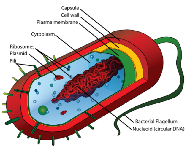
Bacteria are single cellular organisms with a construction oftentimes similar to what is seen in the image above. They have a few main cellular components including a DNA genome, ribosomes for creating proteins, and a large cellular envelope which keeps components inside the cell from escaping and becoming components of the environment in which the bacteria lives.
The eukaryotic cell is quite a bit more complex then the bacterial cell:
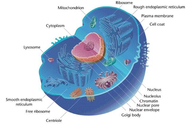
Figure 1: The Eukaryotic Cell
.jpg)
As I am sure you notice (and probably already know!) there are quite a few additional large cellular components in a eukaryotic cell. However the important one for the purpose of this post is the nucleus. Eukaryotic cells have a special compartment where their genetic material is sequestered away from the remainder of the components in the cytoplasm.
...Bacterial Viruses: The Bacteriophage
By now, if you have been reading my blog you are well aware of what a virus is, and you even know about a few different types which are involved in human health. However we mighty eukaryotes are not the only branch of the tree of life that gets infected by viruses. All of the other branches (Bacteria and Archaea) also have their own virus invaders. For bacteria these viruses are called bacteriophages, and they infect bacteria exclusively.
Figure 2: Phage Lifecycle
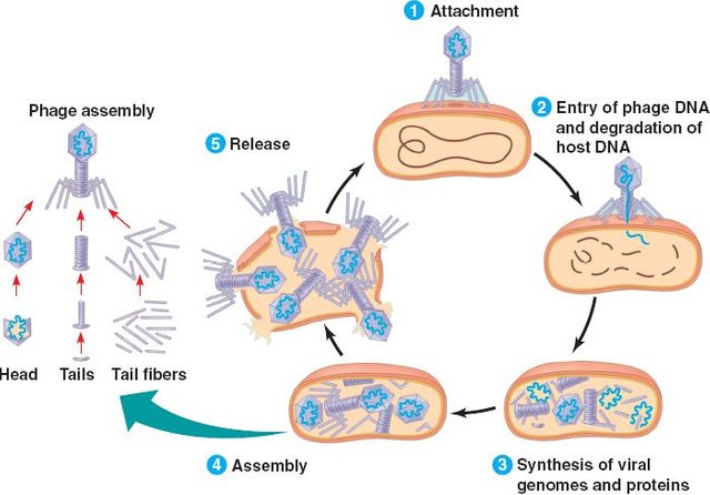
The phage infection life cycle is thought to follow the path outlined in the image above. Initially the virus attaches itself to the exterior of the bacterial cell, and subsequently injects its genetic material into the bacteria. Next, utilizing the hosts DNA replication and protein production machinery the viral genome is replicated and viral proteins (such as its own replication proteins) are produced by the cell.
The newly produced viral proteins get to quick work churning out tons of viral genomes and assembling together new viruses for those genomes to be contained in. Eventually there reaches a point of too many viruses crammed inside the cell (24-48 hours after infection for an E. coli bacteriophage infection for example), that they burst out, rupturing their host cell and killing it. These new viruses spew everywhere and begin their search for a new host to infect, and repeat this cycle all over again.
Dissecting The New Publication
This article is about a bacterial virus named 201φ2-1 (the nerve of the parents of that virus to give it such an awful name!):
If you would like to take a look at what this virus looks like check out this link Here
201φ2-1 infects a bacterium called Pseudomonas chlororaphis.
The researchers were studying the infection of this virus, and to do so they fluorescently labeled one of the proteins in the virus and artificially expressed it in the bacterium, such that they could monitor it by fluorescence microscopy.
The protein they picked was called gp105 (more beautiful science nomenclature for you), this protein is the most abundantly expressed protein upon infection by the virus. Now before the virus infected the bacteria the protein was dispersed evenly through out the cell, however after infection and injection of viral DNA the protein quickly re-arranged into a spherical shaped structure which surrounded the viral DNA. The researchers report that one structure like this was present in every single infected cell, and 10% had two or more of these structures.
They performed some additional experiments and found that proteins could enter the sphere structure, and that the structure rotated in the cell as a whole entity, rather than a collection of protein pieces coming and going.
Next they utilized fluorescent labeling of a variety of other phage proteins and found that proteins responsible for DNA replication processes were localized inside of the viral structure while proteins involved in the creation and assembly of the viral proteins were located outside of the shell. This structure looks and behaves an awful lot like the eukaryotic nucleus!
Evidence for the transition from simple bacterial cells to more complex Eukaroytic cells?
Could the eukaryotic nucleus be a remnant from a viral structure like this one? It seems like a plausible explanation for how eukaryotes might have wound up with such a compartment for their genetic material. We already know that the eukaryotic cell contains what is thought to be the remains of another smaller bacterial cell (called the mitochondria) that was engulfed by a larger cell and continued to live there endosymbiotically.
What do you think? Are our (eukaryotes) cells just a hodgepodge of former smaller cellular (and non cellular) things once living in symbiosis?
At the very least it will be fascinating to see if additional viral structures like this can be observed for other related viruses! Perhaps with more information we will gain more new insights into the evolutionary pathway that life on Earth went on to lead to us!
Additional Sources
- http://science.sciencemag.org/content/355/6321/194
- https://en.wikipedia.org/wiki/Bacterial_cell_structure
- https://en.wikipedia.org/wiki/Eukaryote
- https://en.wikipedia.org/wiki/Bacteriophage
- http://www.fossilmuseum.net/Evolution/Endosymbiosis.htm
All Non Cited Images Are Available Under Creative Commons Licenses
All Gifs Are From Giphy.com and Are Also Available for Use Under Creative Commons Licences
If you like my work, please consider giving me a follow: @justtryme90. I am a PhD holding biochemist with a love for science. My future science blog posts will cover a range of topics in the biology/chemistry fields.
Thank you for your continued support of my work! I appreciate all those who follow me (and those who don't too).
Great article. Maybe one of the many missing links explaining how life developed back then:)
Note that in terms of names, biologists seem not to be better than physicists :)
I think we can't divide our selves into groups for bad nomenclature. Scientists as a whole seem to be terrible with names ;)
I agree! : )
"What do you think?"
Fascinating. However I will not try to answer prematurely a question, which probably so far no one can answer ... But as we know that also mitochondria have been independent organisms before ... everything seems to be possible. :)
Indeed, its honestly an exciting time to be alive for those of us who love science. The amount of knowledge and understanding we are gaining right now is just incredible. Unfortunately here we must continue to wait for more data!
Very interesting and informative. Lots of easily digestible information helping people keep up to date in new developments.
.
ColdMonkey mines [Gridcoin](http://Gridcoin.us) through generating BOINC computations for science.
Thank you @coldmonkey! My goal is to try to do science writing with a bit more background information then seems to be found elsewhere. It's always nice to have a refresher (even when you already know the information), and if you don't know of something then a bit more learning can actually happen. :)