Can Blood Flow In The Brain Can Be Controlled By Light? Research Indicates Yes.
If I shine a light on your brain, will the blood flow patterns in it change?
In this post I will break down some research from a publication in Nature Communications published on January 31, 2017 titled "Light controls cerebral blood flow in naive animals."
In this article the authors were trying to test whether light stimulation protocols similar to those commonly used in opto-fMRI change blood flow patterns in the brains of mice that are not expressing proteins. To better understand their results it is likely best to begin with a bit of information of just what these commonly used techniques are!
Optogenetics and fMRI... Sounds Like more Fancy Science Jargon, What Are They?
Optogenetics
As its name implies, it is a technique which uses light (Opto) to control proteins and genes (genetics) in cells which have been modified to be light sensitive. Yes, there are light sensitive proteins!
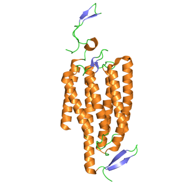
For instance there are ion channels (ion channels are proteins in the membranes of cells that allow/pump ions (think Ca2+ or Na+) into or out of a cell.) Now light sensitive ion channels are those which open in the presence of light, and close in the absence of light. Most of these have been created in labs through research, however there are examples of natural proteins which have this property such as channelrhodopsin which opens in the presence of blue light.
Researchers can use these light sensitive ion channels to change the activity level of certain cells in the brain, by shining a light on them. We can then track how the blood flows in the brain in response to the activation of these activated cellular ion channels through a technique called fMRI.
fMRI
fMRI stands for functional magnetic resonance imaging, and detects brain activity by monitoring blood flow levels. It does this through monitoring the magnetic spin of a "contrast agent." Which is just a compound that allows doctors to see by the MRI technique.
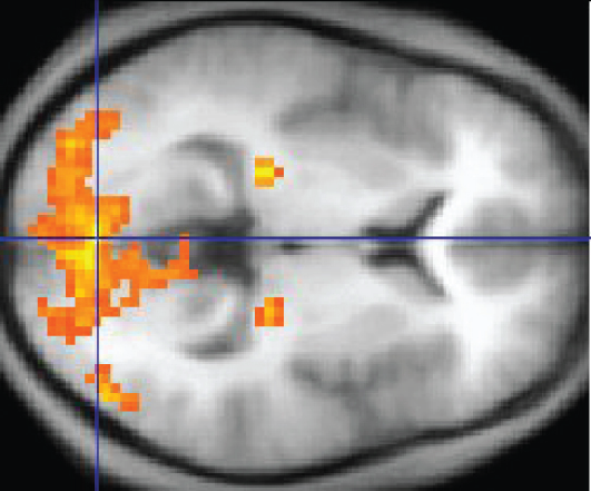
Researchers think that brain blood flow is a good measure of activity because it correlates with the firing of neurons. The more neuronal activity, the more blood flow in that region of the brain. The way blood flow is able to be monitored by the fMRI technique is through use of an oxygen sensitive contrast agent.
The contrast agent they use here is actually hemoglobin! Hemoglobin is the molecule in the body which carries oxygen around for us, and it does so by use of an iron ions in the center of each of its heme subunits. As we discussed above, the fMRI technique is looking for the magnetic spin of a contrast agent, and coincidentally the hemoglobin iron ions have a magnetic spin when they are bound to oxygen but not when they are deoxygenated. So blood oxygen in response to stimulation of the brains activity lights up the fMRI and allows doctors and researchers to see images like this:
So Tell Me About This Article
So as I mentioned above, researchers and doctors use the fMRI technique to monitor brain blood flow, especially in the case with modified cells which are sensitive to light (they contain the light sensitive ion channels), in order to study how the brain functions/responds to the stimulation in certain areas. However one thing that hasn't been studied is the effect that light has on the brains of test cases with out the modified light sensitive proteins (honestly I don't see how this wasn't a control but apparently it wasn't!)
Shining Blue Light On Brains
What they were doing here is shining a blue light (via laser delivered by an optical cable) on portions of mouse brains their setup is shown in (a) below (if you are wondering PMP in the figure stands for polymethylpentene and is just the plastic window they placed overtop of the brain).
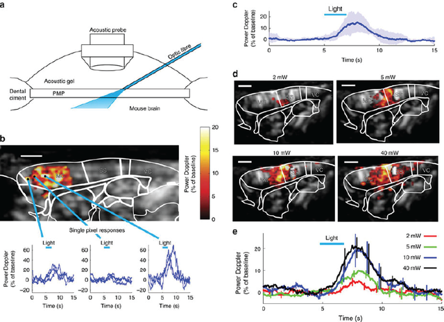
The figure in (b) shows us that they can see increases in blood flow in the brain and can do so on a resolution of a single pixel ( they show curves of differing intensities vs time for a 3 different pixels). They report in (c) the average stimulation of brain blood flow observed between three different mice and in (d and e) they show that the amount of blood flow increase is proportional to the amount of light (the power of the light in Watts) that they blast the brain with.
TL;DR
If you hit a mouse brain with blue light, blood flow in the brain increases even WITHOUT the special light sensitive ion channels. Light it self stimulates brain blood flow.
Why Does This Happen? Dilation Of Arteries
Next researchers wanted to look at the signals that come from the veins and arteries themselves. To do this they used a portion of the brain with a well understood vascular paths called the olfactory bulb. Its the region of the brain responsible for processing smells!
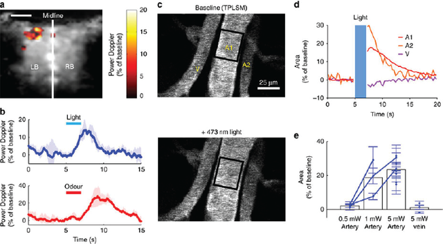
They first verified that hitting the olfactory bulb with the blue light caused a similar increase in blood flow in (a) which they saw it did, so check. They compared the light induced responses with those from letting the mice smell something (olfactory stimulation... it is responsible for processing smells). We can see that in (b) the peak from the light stimulation looks very similar to what they observe from actual functioning of the olfactory bulb (which is pretty neat IMO).
Seeing that they wanted to measure the blood flow levels in the veins and arteries themselves, what they found was that when they hit the brain with the blue light the arteries dilated (aka they got wider) you can see that happen in the two images labeled (c) the ones up top are before being hit with light, and at the bottom after.
They plotted this dilation in (e) and what you see there is that there is more dilation in the artery that correlates to increased amount of light, however veins do not dilate at all (compare the last two bars, the artery dilates a lot, but the vein doesn't do much both with 5 mW of light!)
Other Results
- The light did not stimulate the functioning of the nerves in the brain
- The light did not effect other cells involved in regulating the blood flow arteries (called astrocytes)
- The observed dilation WAS due to a decrease in calcium concentration in the smooth muscle cells surrounding the arteries in the brain.
- The reason previous studies did not observe this was the use of anesthetics on the tested animals. They found that isofluorane anesthesia completely inhibited their signal.
Conclusions
Light delivered at intensities commonly used to trigger fMRI signals leads to dilation of the arteries.
The observed effect is in the same range of amplitude as that evoked by sensory stimulation
The amount of energy required to generate this effect is higher than the threshold used for most opto-fMRI experiments and thus that means of study still remains viable.
More care should be taken for studies that rely upon only blood flow regulation using the BOLD method as the stimulation from light may complicate the interpretation of these studies. Previous studies did not observe the results observed in this study due to their use of isoflurane as an anesthetic.
TL;DR The Whole Post?
We thought special modified cells with photosensitive proteins were necessary for detecting blood flow changes in the brain through the use of light. As it turns out that is not the case and blasting the brain with blue light actually causes activation of regions of the brain and increased blood flow, potentially complicating the interpretation of some old work in this area, but also opening up new avenues for studying the function of the brain.
Sources
- http://www.nature.com/articles/ncomms14191
- https://en.wikipedia.org/wiki/Optogenetics
- https://en.wikipedia.org/wiki/Light-gated_ion_channel
- https://en.wikipedia.org/wiki/Channelrhodopsin
- https://en.wikipedia.org/wiki/Functional_magnetic_resonance_imaging
- https://www.ncbi.nlm.nih.gov/pubmed/24094150
- http://www.medicinenet.com/hemoglobin/article.htm
- https://en.wikipedia.org/wiki/Olfactory_bulb
- http://cshperspectives.cshlp.org/content/7/5/a020388.abstract
All Non Cited Images Are From Pixabay.com and Available Under A Creative Commons License
Any Gifs Are From Giphy.com and Are Also Available for Use Under A Creative Commons Licence
** Nature Communications is Open Access, and Re-use of Information and Images Available Under A Creative Commons Lisence**
If you like my work, please consider giving me a follow: @justtryme90. I am a PhD holding biochemist with a love for science. My future science blog posts will cover a range of topics in the biology/chemistry fields.
Thank you for your continued support of my work! I appreciate all those who follow me (and those who don't too).
I need to get a blue light laser for the next time I write a post! ;)
I'm setting one up right now to help me with my next one. Unfortunately I think this means I need to feed it into my brain... Hmm. I don't think I am qualified to perform such surgery!
Can we try through the nose? Or maybe the ear?! Nope... that wont work, the doctor told me when he looks in one ear he can see right on through and out the other... drats!
Lol :D
It is really interesting, in particular the fact that we may have to revisit many old studies.
Have I got it right if I say that some old results may however not be modified because of the use of a given anaesthetic? But then I guess that there must be other studies relying on other anaesthetic that may be more concerned. No?
Wow, thanks @hilarski! :D
Hi..its off-topic but what's your take on linear radiation for brain tumor? surgery+gamma knife are risky now with surgery in 2013 not effective.
I unfortunately do not know. If surgery was ineffective then radiation is often the next step AFIK, but I am not an MD, so I can not discuss it much I apologise. Perhaps @tfeldman could be of more help?
Don't know much on this topic either. Radiation can be used for treatment/therapeutic (not going to save your life but help relieve neuro symptoms. This occurs a lot in lung tumors as the brain is one of the first places it goes. Might need to talk to a specialist on that one!
@immarojas: my two cents. Gamma rays may not not the best for a tumor as the energy will be spreak all along the ray. I believe hadrotherapy is better as you can use a feature called the Bragg peak to localize the energy deposit better.