The microscopic life observed by my microbiology students.
One of my passions is to teach microbiology at the University. So, some days ago my microbiology students at UCO (Universidad Catolica de Oriente, Colombia) had some training on microscopy in order to study microscopic living organisms, so in this post I want to show some pics of various living creatures under the optic microscopes taken by the students using their mobile phones:
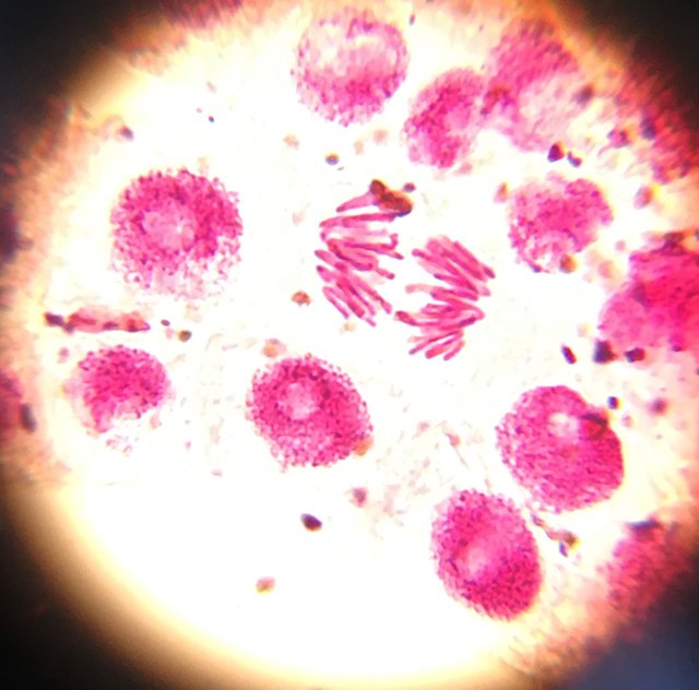
This amazing pic shows the cell division called mitosis in onion cells. Those red colored objects are the nuclei of cells. the filamentous one is showing the cromosomes in a cell division state called anaphase. To obtain this so clear images, the students used a dye called Write, which tinctures the DNA of the nuclei.
.
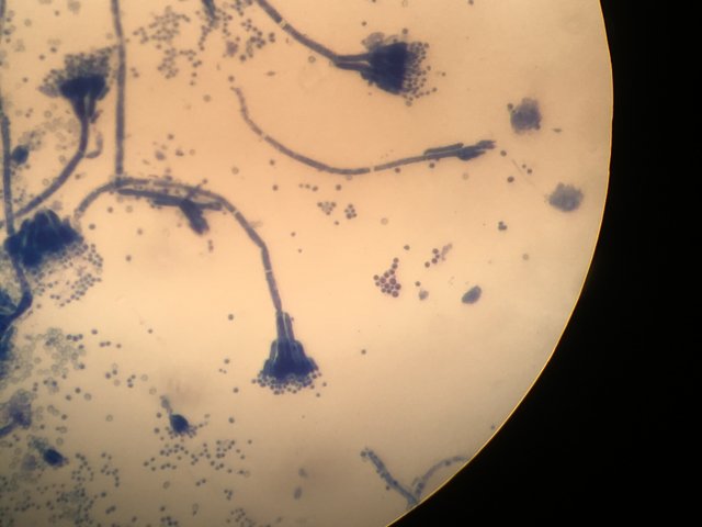
This is a mold called Penicillium, that was grown on bread. This is the same fungus used to produce Penicillin. The spores can be seen very clearly. To obtain this image, students colored the fungus with methylene blue dye.
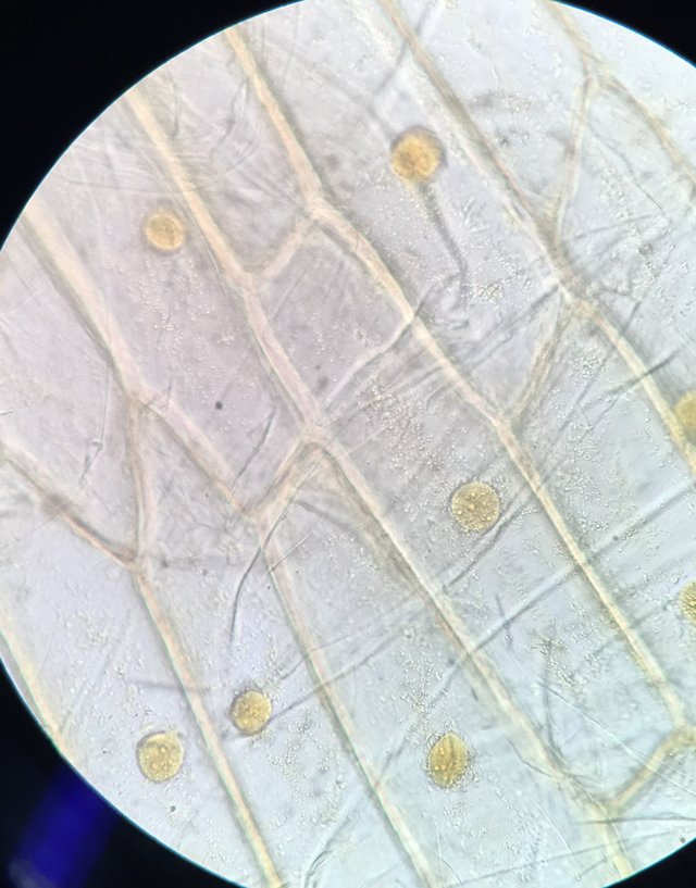
This time, the students found yellow nuclei of onion cells in no cell division. Nuclei were tinctured by using a dye called potasium Iodure.
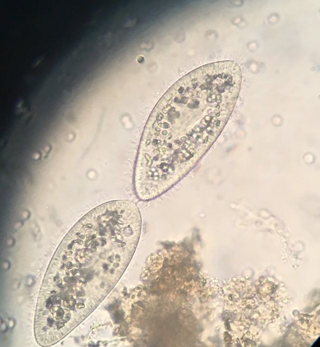
Another wonderfull creatures that students found were those protozoa called Paramecium, obtained from a not so clean water sample.
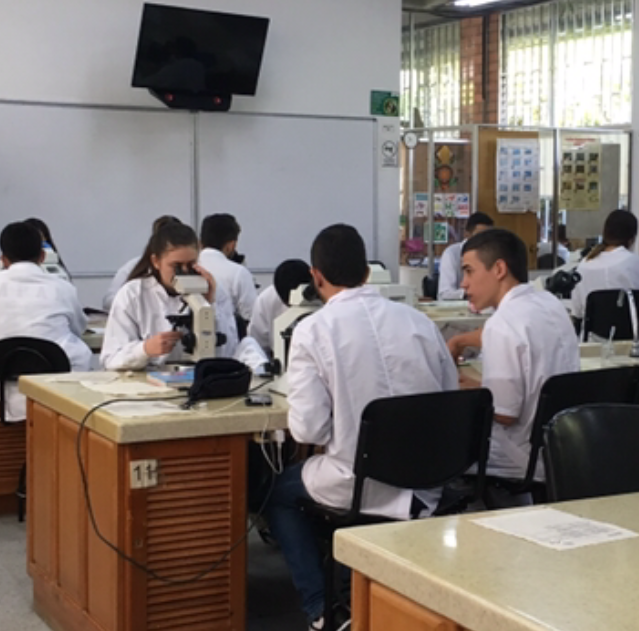
Theese are my dear students working very hard at the lab.
.
Its very satisfying to contribute to the life of the very nice people that in future will direct the destiny of my community.
Finally, I have to say it´s a joy to lern from the findings of my students.