Pulling out a cerebral hydatid cyst located in the brain!
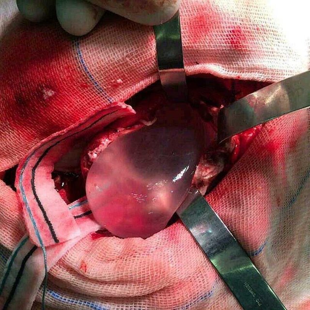
This unique image shows a perfectly round egg-sized hydatid cyst filled with clear fluid within the brain being carefully removed. Brain involvement with hydatid disease occurs in 1-2% of all cases and the common cause is a parasitic infection by Echinococcus granulosus.
The infection is acquired via contaminated food with eggs of the tapeworm. The oncospheres released from the eggs in the bowel enters the portal circulation. Hence the liver is most commonly affected, followed by the lung and other organs such as bones, genitourinary system, bowel and subcutaneous tissues.
Hydatid cysts end up in the brain due to direct invasion of larva that managed and filtered via liver and lung to the brain.
Neurologic examination are nonspecific and depend on the area of the brain involved. They range from mild to full coma and cerebral herniation due to the pressure applied from the space occupying mass. The MRI shows well-defined circumscribed spherical non-enhancing intra-axial cystic lesion that usually lies in the territory of the middle cerebral artery.
Management is surgical, with removal of the entire cyst without rupture using Dowling’s maneuver (instilling warm saline between the cyst wall and the brain) and gently pulling out using retractors to keep some space.