Advocate4Autism #2 - Three aspects of the BRAIN that are different in people with Autism Spectrum Disorder
Over the past decade, there has been extensive neurological research on the differences in brain structure and processing for individuals with ASD. Although we are far from completely understanding the neurological complexities of ASD, we have learned many interesting components of a puzzling world-wide phenomena.
Area #1: Purkinje Neurons
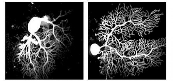
Purkinje neurons are a fascinating component of the intricate neural circuity associated with the region of the brain called the cerebellum. The cerebellum is an area of the brain that is located in the back of the skull, which is often associated with coordination and muscle activity. Strangely, the cerebellum only accounts for 10% of the brain's weight, but it holds over 50% of your brain's neurons. According to recent neurological research, individuals with ASD not only have significantly less purkinje neurons than the average person, but they also have neurons that do not operate in a typical neuronal fashion. For instance, purkinje neurons in people with ASD do not perform a maintenance operation referred to as pruning. Pruning is a neuronal function that serves to eliminate ineffective connections between neurons. In people with ASD, the lack of purkinje pruning creates neural circuitry that is clouded causing strain on coordination and motor skills.
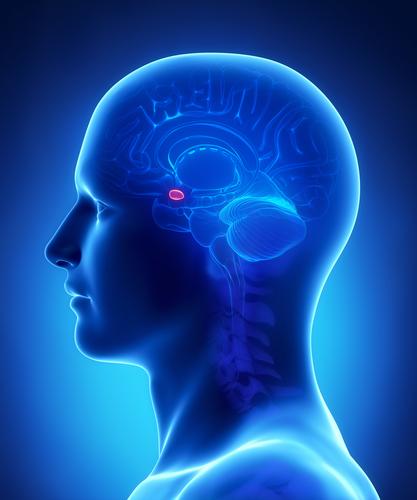
Area #2: Amygdala
.jpg)
The amygdala is a region of the brain that is commonly associated with emotions. According to research conducted by the University of Wisconsin in 2006, the size of the amygdala is smaller in individuals with ASD, which correlates with length of eye contact and general fear of/withdrawal from people. Furthermore, a smaller amygdala is also associated with difficulty recognizing facial expressions and self-regulation.
Area #3: Mirror Neurons
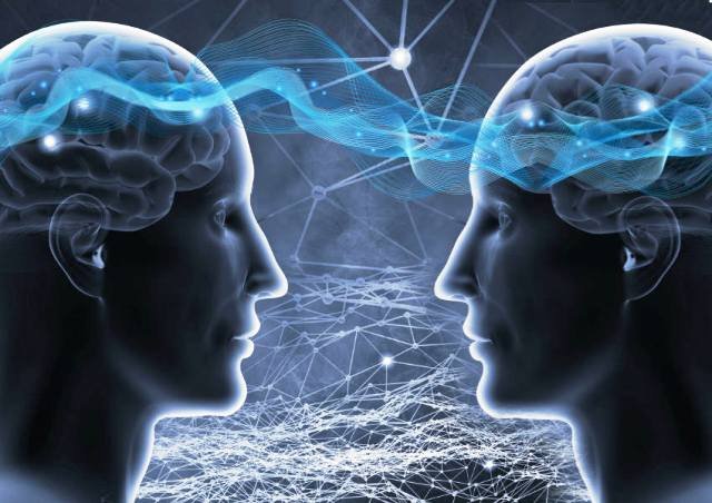
A study by UCLA neuroscientists (Dapretto et al) featuring functional MRI suggests that mirror neurons help people understand the intentions of others – a key component to social interaction. In people with ASD, the research indicates that there is less mirror neuron activity, which significantly muddies an already complex social topography.
If you found this information helpful please click follow and comment below. Thanks for reading my post and helping to spread autism awareness.
Great article!!! Spreading autism awareness and understanding is a great cause. Glad to see people in the steemit community promoting autism awareness.
@johnnycoins
@Johnnycoins Thanks you Sir!