How the centrosome generates an intracellular electromagnetic sphere
The centriole as a rotating device
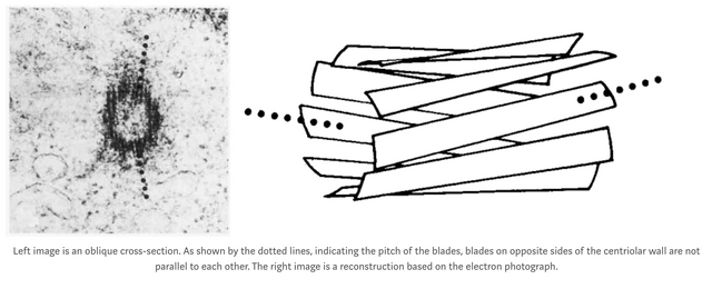
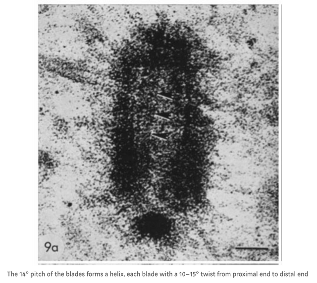
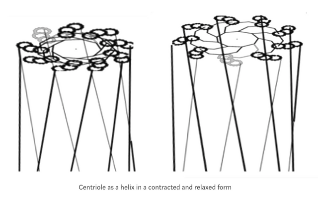
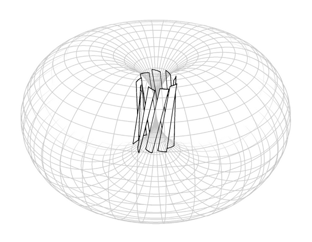
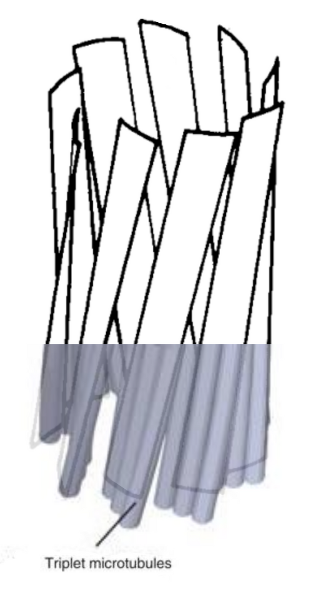
Turns out centriole blades are pitched always[1], centrioles are helical, and that means they pump cytosol along the central axis as they contract.
The rotation is generated by that dynein motor proteins squeeze the whole centriole together, pumping cytosol through it and making it rotate.

The "distal appendages" are distortion in the cytosol, vortices.
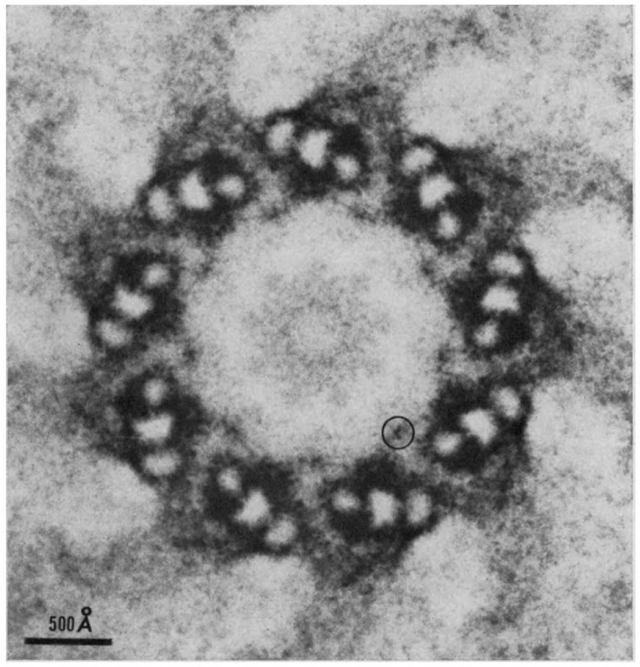
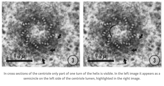
The "vortex" has been observed in the lumen of the centriole in electron photographs[2, 3] is a cytosol vortex, that shows how contraction of the helical centriole pumps cytosol along its central axis.
Synapses
Does the Geometric Design of Centrioles Imply Their Function? (1981)
The three-dimensional structure of the basal body from the rhesus monkey oviduct (1972)
Why the centriole evolved to rotate
Tubulin dimers when excited, electrically charged, produce an electric field that accelerates other charges around it. When these charged tubulin are at rest, as in not moving in space, they do not produce a magnetic field, which is why the centriole has evolved to rotate. The rotation of the centriole, moving the charged particles of each tubulin in each microtubule triplet through space, perturbs the surrounding environment and generates a magnetic field perpendicular to the direction of the particles motion, and when the magnetic field at each point in the circumference of the rotation is added together, it forms a torus.
The centriole as a turbine-pump, and the centrosome orbital system
The centriole is a turbine[1] that also generates the flow of cytosol that powers its rotation, and does so through squeezing itself together, like a pump, causing it to rotate as well as to pump cytosol downwards along the central axis and out at the proximal end, propelling the centriole in a forward direction.
The forward movement of each centriole of the centrosome, combined with that both also generate an electromagnetic field through rotation of the barrel structure[2] composed of electrically charged tubulin dimers, will cause them to orbit one another, both centrioles partaking in a dance where they spiral around one another, generating another level of rotation that gives rise to an electromagnetic sphere that is the combination of the EM field of both centrioles and the orbital movements they partake in.
Synapses
Note on the observation of turbulence in electron photographs of centrioles
The centrioles as molecular dynamos need to both rotate, and oscillate.[1] If that is not turbulence, then what part of a dynamo would it be? "subdistal appendages" that are attached to a rotating body, how does that work? To begin with, would they not increase drag and slow the rotation, be a structure that is in opposition to force from the contraction that leads to rotation and generates the magnetic field?
These "appendages" for example, modelled in this meme from 2018, they are the opposite direction to the blades, they are either a) a drag on the rotation, counter to what is expected to have evolved for a dynamo, or b) cytosol being distorted from the pumping that causes rotation.
With closer examination, it is impossible for those to be "appendages" if the centriole is to be able to rotate, which means a) the centriole does not rotate or b) they are not appendages, but cytosol whirls.
Synapses
On cytosol whirls from the centriole as a turbine-pump driven by contraction of motor proteins


How does a cytosol vortex behave at the nano scale?
"In the search for the miraculous, we find that its often right before our very eyes but we fail to see it. " - Jason L. Silva
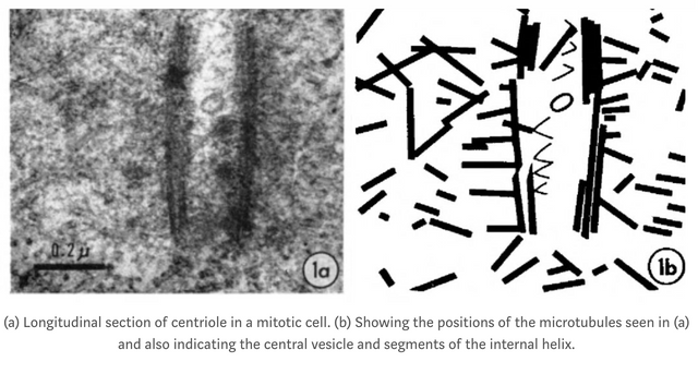
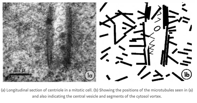
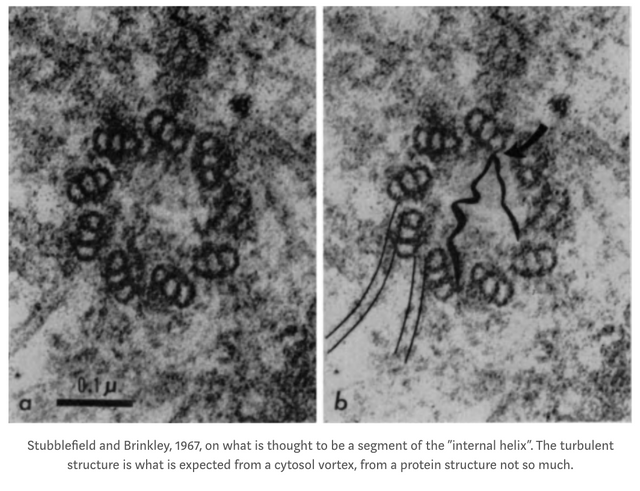
Perhaps a cytoplasm vortex at the nano scale differs from water vortices at the macro scale that people are used to observing, so that evidence of them goes unrecognized for 40 years? The "internal helix" in centrioles was first observed in the 70s, or, the first mention I have seen at least is from 1967.
Water molecules have a width of 0.275 nanometers, and the centriole is 500 nm high and 250 nm wide, and the width is dynamic since the entire centriole as a turbine-pump contracts to pump cytosol through the central axis also causing it to rotate similar to a turbine.
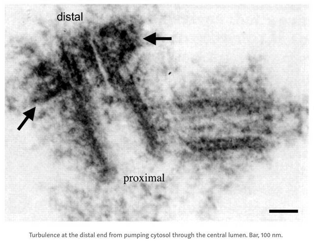
In most vortices a strong sub pressure (vacuum) is generated in the center, and concentrates substances, seen as a string of particles following the sub pressure. In the cytosol vortex within the centriole as a turbine-pump, the vacuum string is about the width of a microtubule wall (see middle and bottom image), roughly 5 nm, the same width as 18 water molecules if each has a diameter of 0.275 nm. Because the centriole itself is rotating, the sub pressure string in the cytosol vortex, the part of the vortex that is discernible in electron photographs, is spiralling down the central axis of the centriole, also taking a more turbulent path at times (see bottom image. )
Delete comment
That's how the web works since decades now, information wants to be free, it copies itself, like cells divide or genes spread when mammals mate. Google actually crawls the web since two decades and eats information, that I love. Re: your scam, does not seem very profiting, why would you choose to work with that even when there are more rewarding endeavours?
Sorry dude but my account was hacked luckily i retuned it back!
interesting pattern it has some unique properties too,,,,
thanks
There are several questions for your observation, the first one is did distal appendage of centriole attached to centrosome during mytosis? The electric microscope picture of centrosome you cited might from cells that are not the M phase of cell cycle, in which the centrosome is anchored with cytoskeleton, and centriole may not rotate when cell is not dividing, also centrosome itself may need sometime to duplicate.
Secondly centrosome distal appendage only attached to one centriole of the centrosome, so is it possible that it functions as the anchorage structure of the centrosome? I believe centrosome position is fixed in nondividing cells, so perhaps in G0,G1,S,G2 phase of cell cycle, centrosome is less dynamic than in the M phase, one of the centriole is anchored with cytoskeleton, and not rotating? Nevertheless the centriole without distal appendage may still rotates when centrosome is anchored to cytoskeleton.