Basic Emotions and the Anatomy of Face Muscles
Basic Emotions and the Anatomy of Face Muscles
/GettyImages_170955996-56a794ec5f9b58b7d0ebe594.jpg)
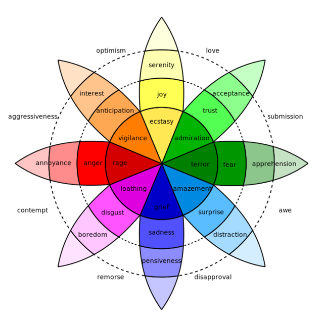
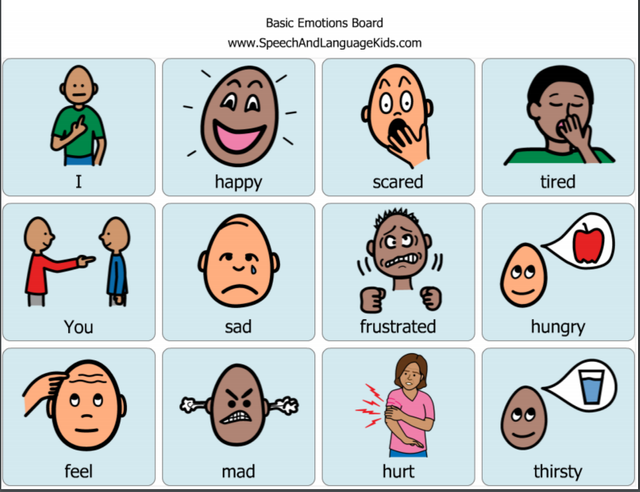
Humans have a pallet of feelings that we inherit through our genes. So every person in the world, regardless of culture, has access to the same emotions. Emotions are sometimes clearly culturally shaped and that they cannot be read off easily from body language. It is not possible to set up a kind of universal for all countries in the world and to clearly interpret the true feelings of others without any background knowledge.
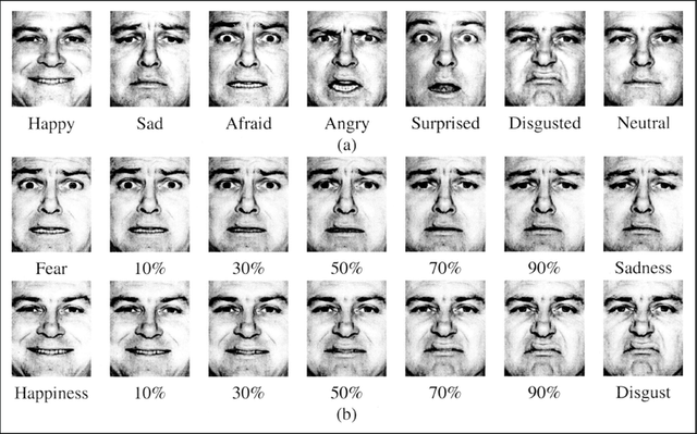
The facial expression of the 7 basic emotions is independent of culture worldwide. Anger and disgust look exactly the same with the natives in some other countries. These facial expressions are the only ones that can be clearly read out of context and none of the other expressions.
Expressions for contempt use the same muscle groups on the face, only that they are unilaterally contracted when contemptuously. The corner of the mouth rises on one side, the nose wrinkles on one side, etc. The detection rate of emotions is lowest for fear and surprise. These two basic emotions are very often confused with each other because they trigger a similar facial expression, the lifting of the eyebrows. If you are surprised, your forehead is evenly furrowed, but if you are afraid, they also cramp over your nose so that the furrowing doesn't look quite as even.
Anger is often confused with disgust, because both feelings occur in similar situations where hatred and aversion meet. With a little more precise knowledge of facial expressions, you can easily distinguish them, because disgust wrinkles your nose while the eyebrows hardly move. In contrast, when angry, the eyebrows are drawn together, while the nose is not furrowed.
Face Muscles
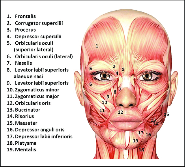
Facial muscles is called a group of muscles in the area of the face which in the present above the skin radiate and by their motion the physical expression of emotions and moods. Facial muscles are not connected to joints like most others, but they mostly insert into the skin without an attachment tendon. They have no fasciae, they only serve to move the skin of the face.
When a person's eyebrows are jerked up, a number of small muscles are responsible for it. In order to be able to describe such a movement, it would now be necessary to describe which muscle is doing what. That would be too much of a hassle.
Classification of Facial Muscle
FOREHEAD
Occipitofrontalis muscle
The occipitofrontalis muscle belongs to the epicranii muscles. It has two muscle bellies at opposite poles of the skull, which are connected by the galea aponeurotica.
The venter frontalis of the muscle has its origin at the supraorbital margin of the frontal bone and in the area of the glabella. Its fiber strands radiate into neighboring facial muscles, including the procerus muscle, the corrugator supercilii muscle and the orbicularis oculi muscle while venter occipitalis has its origin in the linea nuchae suprema of the os occipitale and on the os temporale.
Temporoparietalis muscle
The temporoparietalis muscle is a thin muscle running on both sides of the lateral skull which belongs to the epicranium muscles. The posterior part of the temporoparietalis muscle is also referred to as the superior auricular muscle and counts among the ear muscles.
EYE
Orbicularis oculi muscle
The orbicularis oculi muscle is a muscle in the area of Orbitaoffnung, circulating the eye and the palpebral fissure.
Corrugator supercilii muscle
The corrugator supercilii muscle is a small, pyramidal muscle in the area of the eyelid. Its fibers run below the occipitofrontalis muscle and partially below the orbicularis oculi muscle obliquely cranially and laterally. There they shine into the skin of the side eyebrows where they find their roots.
Depressor supercilii muscle
The muscle depressor supercilii located at the processus frontalis of the maxilla and from the arcus superciliaris of the os frontale. The muscle has two heads, between which the angular artery and vena pass. Its fibers run steeply cranially. There they shine into the skin of the medial eyebrow, where they begin.
NOSE
Nasalis muscle
The nasalis muscle is a two-part muscle in the area of the nose. Pars transversa originates from the maxilla above and slightly to the side of the incisive fossa. Its fibers run cranially and medially and insert on both sides in a thin aponeurosis that covers the bridge of the nose. Some fibers radiate into the aponeurosis of the procerus muscle. Pars alaris has its origin in the incisive fossa of the maxilla somewhat medial and caudal to the origin of the transverse pars on the bone parts above the root of the 2nd incisor. From there, the fibers pull into the cartilage of the nostril and to the tip of the nose.
Depressor septi nasi muscle
The depressor septi nasi muscle has its origin in the incisive fossa of the upper jawbone. Its fibers run medially and cranially. There they radiate into the cartilage of the nasal septum and the nostril, where they find their attachment.
Procerus muscle
The procerus muscle has its origin in the fascia above the insertion of the nasal bone. Its fibers run steeply cranially, there they shine into the skin of the glabella, where they find their base. Some strands of fibers mix with those of the frontal vent of the occipitofrontal muscle.
Levator labii superioris alaeque nasi muscle
The levator labii superioris alaeque nasi muscle is a superficial muscle in the anterior cheek area or on the slope of the nose that lifts the upper lip and the nostril. Has its origin at the lower edge of the eye socket or on the frontal side of the upper jawbone. It's fibers run along the nose slope after caudal to the upper lip and the nostrils. Some fibers of the muscle radiate into the orbicularis oris muscle.
MOUTH
Orbicularis oris muscle
The orbicularis oris muscle is a muscle that surrounds the mouth opening in a circle and closes the lips. The origin of the orbicularis oris muscle, the anterior surfaces of the alveolar ridges of the upper and lower jaw. Fibers run in concentric circles around the cleft lip. In the area of the corner of the mouth, the fibers of the orbicularis oris muscle combine with the fibers of the buccinator muscle and other facial muscles to form the so-called modiolus.
Depressor anguli oris muscle
A thin muscle in the area of the mouth that pulls the corners of the mouth downwards. Has its origin at the mental tubercle and at the lower edge of the mandible. The fibers run cranially to the modiolus at the corner of the mouth.
Risorius muscle
A superficial muscle in the area of the mouth and cheek that pulls the corners of the mouth to the side. The risorius muscle has its origin in the skin of the cheek and on the fascia of the masseter muscle. The fibers pull medial to the modiolus at the corner of the mouth.
Mental muscle
Also a superficial muscle in the area of the mouth that tightens the chin skin. Musculus mental has its origin at the mandible in the incisive fossa on the alveolar wall of the second mandibular incisor. The fibers pull caudal to the chin skin.

Levator labii superioris muscle
A superficial muscle in the front of the cheek or on the slope of the nose that pulls the upper lip upwards. The levator labii superioris muscle runs in three fiber strands or muscle heads.
The medial fiber strands are also known as the angular caput. This part of the muscle is identical to the levator labii superioris alaeque nasi muscle. It has its origin in the frontal process of the maxilla and runs obliquely downwards to split further caudally into two fibers. Some of the fibers insert into the alar cartilage and the skin of the nostril the other part of the fibers radiates into the lateral part of the upper lip and mixes there with the fibers of the orbicularis oris muscle. The middle portion of the fiber strands arise from the lower edge of the orbit just above the infraorbital foramen. The fibers converge in their course in the caudal direction and radiate into the muscle substance of the upper lip between the canine and the angular head. The lateral fibers form the zygomatic head which is identical to the zygomaticus minor muscle arise from the bony surface of the os zygomaticum behind the suture zygomaticomaxillaris and pull downward and medial to the upper lip.
Zygomaticus major muscle
The zygomaticus major muscle has its origin at the zygomatic bone, more precisely at the anterior end of the processus temporalis, behind the sutura zygomaticotemporalis. The fibers run medially and caudally to the modiolus at the corner of the mouth and to the upper lip.
Zygomaticus minor muscle
The zygomaticus minor muscle originates from the zygomatic bone immediately in front of the zygomaticomaxillary suture. The fibers run medially and caudally to the upper lip.
Buccinator muscle
The buccinator muscle found on the outer surfaces of the alveolar processes of the upper and lower jaw, for example in the area of the three molars, as well as on the crista buccinatoria. Additional fiber strands arise dorsally from the front of the pterygomandibular raphe which separates the muscle from the superior pharyngeal constrictor muscle.
Depressor labii inferioris muscle
The depressor labii the inferior located at the lower border of the mandible to the oblique line between symphysis and mental foramen. Here the muscle continues the fiber course of the platysma. Its fibers run steeply superiorly and medially to the lower lip, where her approach found in the skin of the lower lip. Some of them mix with the fibers of the orbicularis oris muscle.
Levator anguli oris muscle
The levator anguli oris muscle located in the canine fossa of the upper jawbone just below the infraorbital foramen. Its fibers run caudally and laterally to the corner of the mouth. There they mix in the modiolus with the fibers of the zygomaticus major muscle, the depressor anguli oris muscle and the orbicularis oris muscle.
