Dental Basics || Teeths || Dentistry || Education || Wednesday || 28.10.21
Assalam o alaikum to all of my lovely steemit members.
Today is wednesday and my focus for today is gonna be based on Dentistry.
You all must have known by now that I am doing BDS (Bachelor of Dental Surgery) and currently in 2nd year. I get a lot of assignments and projects regarding dentistry so today I thought why dont I share my project with you guys too.
When I was in 1st year of BDS we got ample of projects but I dont exactly remember all of them. Few of the projects I remember were:
Wire work
Plaster slab
Partial denture
Waxing
Bridge placement
Proportioning and mixing of Impression Materials
Wire work was based on soldering and brazing of metal wire. It was performed to teach us about the basics of orthodontics. We got a good knowledge about braces placement and hamdling of braces along with whole of the braces procedure in this project.
Plaster slab was basically taught to know about mixing and proportoning ratios of impression materials. Other than this, it had nothing to do with our dental practice.
Waxing and de-waxing are performed on a denture (denture is basically a model of tooth/teeths which is usually made if plaster of paris). One sole reason of waxing is to acheive a perfect impression of a tooth/teeths.
Bridge placement again is fixing an abnormality in a denture. Its is basically replacing a couple of theeths or more than two, with a new ones which are made in laboratory and then fixed into patient's mouth in a hospital by the dentist.
Last experiment in my first year of BDS was Proportioning and mixing if dental materials. In this experiment techers basically teach us about the ratios of different compartments needed to get a perfect impression.
Impression can be taken through alginates, impression compounds, agar etc.
Now coming to the Second year projects.
I am recently working on Tooth Carving. Its a very wide procedure and I am gonna discuss in detail about it below.
TOOTH CARVING
We have total four quadrants in our mouth. Each quadrant has 8 teeths. We got an assignment to carve these 8 teeths i.e one full quadrant, on a carving wax block.
Carving wax block looks like this:
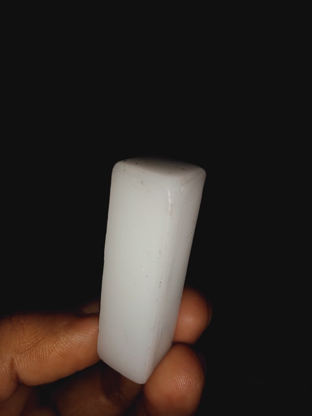
It is rectangular in shape. It is tough and hard and is used for making tooth forms in the study of Dental Anatomy.
Carving wax block is easily available in any of the pharmacies.
It is not much cheap. I bought 10 blocks for 170 rupees only.
In order to carve teeths from a carving wax block, we will need a dental wax carver also.
Dental wax carver is also available in any of the dental markets.
We had to carve following 8 teeths with this carving wax block:
- Maxillary Central Incisor
- Maxillary Lateral Incisor
- Maxillary Canine
- Maxillary first Pre-Molar
- Maxillary second Pre-Molar
- Maxillary first Molar
- Maxillary second Molar
- Maxillary third Molar
Among these eight teeths I've carved only four uptil now. And remaining four I am gonna carve next week so I will share remaining for in the next week's post.
In this post I am gonna share all the details of the four teeths which I have carved i.e:
Maxillary Central Incisor
Maxillary Lateral Incisor
Maxillary Canine
Maxillary first Molar
I have carved all four of the teeths mentioned above.
Now lets start with the first teeth!
Maxillary Central Incisor
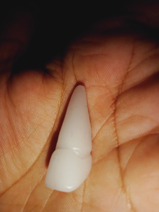
Maxillary central incisor is a tooth present in the front upper jaw also known as maxilla . This tooth is present at the very front so it is usually the most visible of all teeths in the mouth.
Location:
It is located mesial, which is near to the midline of the face, to the maxillary lateral incisor.
Function:
Function is same as all the othet incisors. Their function is for shearing (tearing flesh) or cutting food during mastication (mastication is another word for chewing which also means physical digestion of food which takes place in the oral cavity). There is only a single cusp on each incisor tooth which is called an incisal ridge or can also be termed as incisal edge.
Origination:
Incisors usually originate and appear to develop at 14 weeks in utero for the deciduous (baby teeths or milk teeths) set and 3–4 months of age for the permanent set(adult teeths).
Dimensions:
Length of crown: 9 mm
Length of root: 13 mm
Diameter of crown: 6.5 mm
Diameter at cervix: 5 mm
Below here I am attaching a picture of Maxillary Central Incisor which I carved on a wax block:
Maxillary Lateral Incisor
Maxillary lateral incisors appear in form of a pair in upper jaw (maxillary area).
Location:
Maxillary lareral incisors are located laterally, i.e away from the midline of the face, from both maxillary central incisors of the mouth and medially, i.e towards the midline of the face from both maxillary canines (maxillary canines are also discussed later in this post).
Function:
As I mentioned above, all the incisors exhibits same function. Their function is for shearing (i.e tearing) or cutting food during mastication( also known as chewing and can also be termed as physical digestion which occurs in oral cavity and is performed by teeths).
There are usually no cusps found on the Maxillary Lateral Incisors but there's a rare condition known as "Talon cusps". They are most prominent on the maxillary lateral incisors The surfac of the tooth which is used in eating is called an incisal ridge or can also be call as incisal edge.
Origination:
There irigination is almost same as Maxillary Central Incisors, but there are some minor differences between the deciduous teeths (baby teeths or milk teeths) Maxillary Lateral Incisor compared to that of the permanent Maxillary Lateral Incisor. The Maxillary Lateral Incisors always obstructs the position of the Mandibular (lower jaw) Lateral Incisors.
Dimensions:
Length of crown: 9 mm
Length of root: 13 mm
Diameter of crown: 6.5 mm
Diameter at cervix: 5mm
Here I am adding a picture of Maxillary Lateral Incisor which i carved on a wax block:
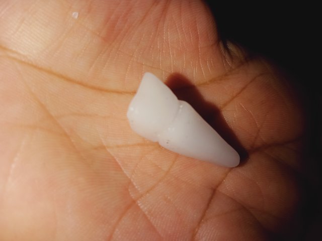
If you compare the pictures of Maxillary Central Incisor and Maxillary Lateral Incisor then they would almost be same because obvioulsy their structure is same and so are functions. But if you look closely, there is a dimension difference in between both of them.
Maxillary Central Incisor is a bit bigger than Maxillary Lateral Incisor.
You can compare the dimensions if you are looking up to a difference in both of them.
Maxillary Canine
The Maxillary Canine is the tooth located laterally i.e away from the midline of the face, from both the Maxillary Lateral Incisors of the mouth but mesial from both maxillary first premolars(Pre molar will also be discussed soon in my upcoming posts). The maxillary and mandibular canines both are collectively called the 'cornerstone of the mouth'. The reason behind is because they are both located three teeth away from the midline of the face and
also separates the premolars from both of the types of incisors.
Location:
Canines are located in such position that they reflect their dual function. Their dual functions includes complementing both the premolars and both types of incisors during mastication, commonly known as chewing (as mentioned above also).
Function:
Most widely, the most common action of the canines is tearing (shearing) of food. The canines most usually occludes in the upper gums few millimeters above the gum line. The canine teeths have the ability to bear the enormous lateral pressure which caused by chewing (also mastication). There is only a single cusp on canines. There structure is moulded in such form that they resemble the prehensile teeths which are most commonly found in carnivorous animals.
Dimensions:
Length of crown: 10 mm
Length of root: 17 mm
Diameter of crown: 7.5 mm
Diameter at cervix: 5.5 mm
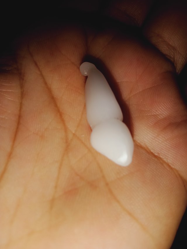
Below here I am attaching a picture of Maxillary Canine which I carved on a wax block:
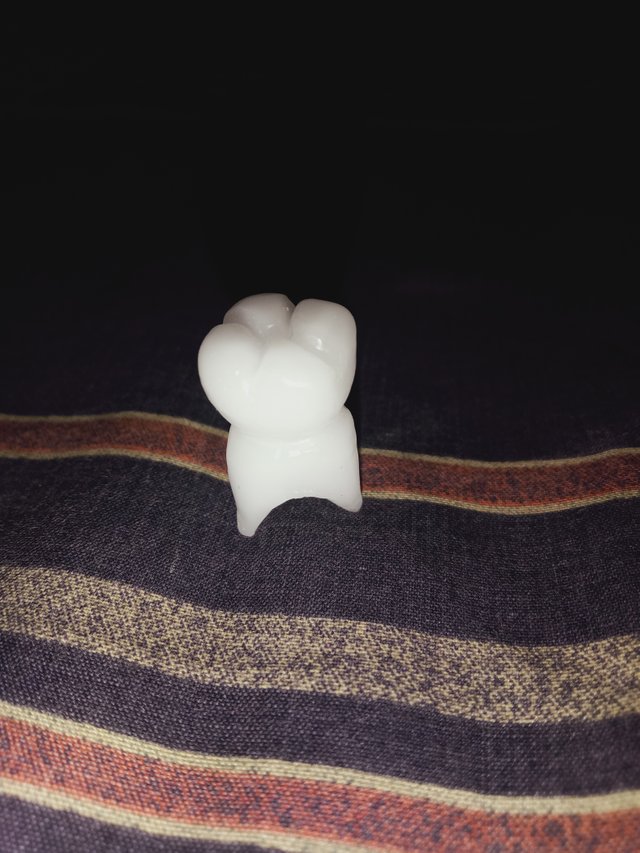
Maxillary First Molar
Maxillary first molar is the sixth tooth of the eight teeths of second quadrant which is present near to the end of the oral cavity.
Location:
It is located laterally i.e away from the midline of the face, from both the maxillary second premolars of the mouth but mesial i.e toward the midline of the face, from both the other maxillary second molars.
Functions:
The function of maxillary first molar resembles to the second and third molars. Their pricipal action during the mastication (commonly known as chewing) is Grinding of food.
Dimensions:
Length of crown: 7.5 mm
Length of root: 12, 13 mm
Diamater of crown: 10 mm
Diameter at cervix: 8 mm
Below here I am attaching a picture of Maxillary first molar which I carved on a wax block:
This is all about dental education I had to share with you all today.
I hope this was helpful and beneficial for you guys.
If you like these kinda educations stuff please do inform me. I would love to share my other dentistry related adventures with you guys.
Please show support if you like it.
Thankyou.
I'll be back with another post soon.
Till then, take care of yourself.
Stay happy.
Stay blessed.
Good Bye!
Wonderful you have done your post very well and your post is commendable
Thankyou.