NEURONS
Sourcre:pixabay
The human central nervous system contains more than 100 billion neurons.About 40% of human genes participate in the formation of Neurons.The primary functions of Neurons is integration and transmission of nerve impulses. The Neurons, endocrine and nervous systems together form the control system of the body.
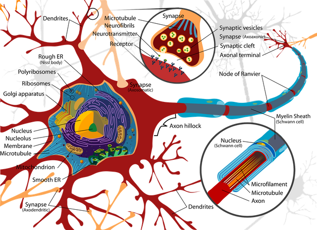
The neuron is the structural and functional unit of the nervous system. It is similar to other cells in the body by having nucleus and other organelles found in the cytoplasm. However, neurons differ from other cells of the body by the presence of axion, dendrites, nissl granules and neurofibrillae.They also lack centrosome, thereby losing the power of division. Unlike other cells, neurons contains and secrete neurotransmitters.
There are three different ways of classifying neurons. There includes:
i, depending on the number of poles it has.
ii, depending on it functions.
iii, depending on the the length of the axion.
Depending on the numbers of poles, neurons can be classified into three types. Namely:
a, Unipolar : which has one pole and from that single pole arises the axion and dendrites. This type of neurons are found in embryonic stages in human.
b, Bipolar :which has two poles, the axion arises from one of the poles while the dendrites arise from the other pole. This type of neuron can be found in the retina.
c, Multipolar: this type of neurons has many dendrites, and the axion arises from one pole while the rest give rise to dendrites.
Based on their functions, neurons can be classified into two.
i, Motor or Efferent neurons :this neurons transmit information from the central nervous system to the effector organs or glands. Motor or Efferent neurons usually have long axion and short dendrites.
ii, Sensory or Afferent neurons :this are neurons that carry information from the peripheral nervous system to the central nervous system.
Based on the length of their axons, neurons can be classified in to two. They are
i, Golgi type I
ii, Golgi type II
Golgi type I usually have long axons and their cell body is situated in the central nervous system. Their axons also reaches remote peripheral organs.
Golgi type II neurons has short axons and are present at the cerebral cortex and spinal cord.
Neurons are made up of the cell body, axon, dendrites and the nerve terminal
The cell body also called the soma varies in shapes and sizes such as stellate, round and pyramidal. The nerve cell body main function is the structural integrity of the neuron.
The cytoplasmic contents of the nerve cell body includes the nucleus, nissl body, neurofibrillae, mitochondria and golgi apparatus.
Each neuron has one centrally placed nucleus in the soma, a prominent nucleolus which contains ribonucleic acids but lacks centrosome thereby losing it power of division
The nissl granules or bodies are small basophilic granules or membrane bounded cavities found in clusters or clumps in the cell body. They are present in cell body and dendrites but absent in the axon and axon hillock. Nissl bodies are composed of ribonucleoprotein. They synthesize proteins of the neuron which are transported by the axonal flow. When the demand for protein synthesis increases, the nissl bodies overwork and may although disappear. They reappear following recovery of neuron from fatigue, anoxia or injury.
The neurofibrillae are thread like structures that are present all over the nerve cells or neurons. They consist of microtubules and microfilaments.
Mitochondria are present in soma and axon and forms the power house of the neuron where Adenosine triphosphate(ATP) are produced.
The golgi apparatus does the work of processing and packaging of proteins into granules in the neuron or nerve cell.
The dendrites are branching extensions of the soma. The dendrites of the cerebral cortex shows knobby projections called dendritic spine.Dendrites maybe absent, single or multiple depending on the type of neuron. The dendrites conduct impulses towards the cell body. It also generates local potential but not action potential.
The axon arises from the axon hillock and is one in a neuron. Axon carry impulses away from the cell body and cannot synthesize it own proteins but depends on the soma for protein synthesis. It branches only at the terminal end called synaptic knob or terminal button. The axon is specialized to convert electrical signals into chemical signals.
Some neurons has axon with myelin sheath while some lacks the myelin sheath. The myelin sheath are concentric layers of proteins alternating with lipids.
Nerve fibre that are insulated with a myelin sheath are called myelinated nerve fibres while those that are not are called non-myelinated nerve fibres. Myelinated nerve fibres are 100 times faster than non-myelinated nerve fibres when transmitting impulses.
Outside the central nervous system, myelin sheaths are produced by the schwann cells, while in the central nervous system they are produced by the oligodendrocytes. The myelin sheath are not continuous, they are absent in the node of ranvier.
Myelinogenesis is the formation of the myelin sheath around the axon of peripheral nerve cells which normally starts at the fourth month of intrauterine life and complete few years after birth.
Myelin sheaths makes the propagation or transmission of action potential very fast. It also acts as an insulator to prevent leakage of electrical impulses during transmission.
References
Modern Biology by Sarojini T. Ramlingam-ISBN 9781754370
College Biology by Idodo Ume-ISBN 9788052207
Essentials Of Medical Physiology by K. Sembulingam, Prema Sembulingam-ISBN 97881-8448-704-6
Textbook Of Physiology by Gabriel C. Ezeilo-ISBN -13:978-0-19-5665-7