The small intestine
THE SMALL INTESTINE
.jpeg)
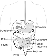
The part of the alimentary canal extends from the pylorus of the stomach to the proximal end of the large intestine: In man it is about 20ft long when fully relaxed, but in life its smooth muscle is in State of tonic contraction and the length is usually about 7 feet. The processes of digestion which-are started in the mouth and stomach are carried to completion in the intestine with the aid of secretion from its epithelium and from accessory glands. The absorption of most of the nutrients take place through the epithelium of the small intestine.
They are carried away in the blood and lymphatic vessels. The small intestine is divided into three regions namely, the jejunum, the duodenum and the ileum. There are slight differences between these regions with a gradual transition between them, but the general arrangement of tissues comprising the wall is in principle the same throughout its length. There are layer; the serosa, the muscularis, the submucosa and the mucosa. The outer serous consists of a membrane of connective tissue which is continuous with the mesentery and the lining of the abdominal cavity. These membranes collectively known as peritoneum enclose the peritoneal cavity. The mesentery is a double membrane of connective tissue at the midline of the posterior abdominal wall. It supports the intestine but at the same time allows them considerable freedom of movement. The muscularis contains two layers of smooth muscle. The outer layer lying immediately beneath the serosa has a longitudinally arranged fibres and the inner layer has circularly arranged fibres. Between the two muscle layers there are many ganglion cells and a network of nerve fibres termed the mesenteric plexus of Auerbach. Nerve fibres, from the plexus innervate both: muscle layers. The next layer, the submucosa consists of adipose connective tissue. In the duodenum the submucosa contains the secretory alveoli of glands. Another network of nerve fibre and ganglion cells in the submucosa is known as submucosa plexus of Meissner. The lamina properial divides the submucosa plexus of mucosa. This is a fibroblastic connective tissue containing loosely arranged smooth muscle fibre of the muscularis mucosae.
.jpeg)
https://www.google.com/search?q=the+small+intestine&oq=the+small&aqs=chrome.0.69i59j0l2j69i57j69i60.3964j0j4&client=ms-android-hmd&sourceid=chrome-mobile&ie=UTF-8#imgrc=INYIMLZNKKC3CM
The muscle cells are innervated by axons
meissner's plexus. The lamina properia also contains many blood vessels and lymphatics. The basement membrane of the epithelial cells of the mucosa is formed by fibres of the lamina properia. Many of the epithelial cells are adapted for special functions: some are absorptive and have brush borders others are secretory and contain granules or appear as goblet cells.
ORGAN BATH FOR INTESTINAL OMTILTY EXPERIMENT INTRODUCTION
Smooth muscle ( involuntary or visceral muscle) differs from striated ( voluntary)_ muscle by the slowness of its contraction and relaxation and by its property of inherent contractility.
Thus when a piece of isolated smooth muscle tissue is suspended in a suitably warmed, aerated saline solution (e.g. Tyrone's solution) it exhibits tone and gives rhythmic contractions. Within the body this activity is normally modified by autonomic nerves supplying the tissue. In vitro, the action of the nerves may be mimicked by the addition to the bathing medium of the humoaal substance normally released at the nerve endings.
Reference
Nosek, Thomas M. "Section 6/6ch2/s6ch2_30". Essentials of Human Physiology. Archived from the original on 2016-03-24.
human body | Britannica.com
a b c DiBaise, John K.; Parrish, Carol Rees; Thompson, Jon S. (2016). Short Bowel Syndrome: Practical Approach to Management. CRC Press. p. 31. ISBN 9781498720809.
Tortora, Gerard (2014). Principles of Anatomy & Physiology. USA: Wiley. pp. 913. ISBN 978-1-118-34500-9. ..its length is about 3m in a living person and about 6.5m in a cadaver due to loss of smooth muscle tone after death.
Standring, Susan (2016). Gray's Anatomy. UK: Elsevier. p. 1124. ISBN 978-0-7020-5230-9. ..and has a mean length of 5 metres (3 - 8.5 metres) when measured intraoperatively in the living adult (Tietelbaum et al 2013).
Graet write up
Keep it up..
Great research
Nice one 🌹
Great one dear.....I love this
What a nice Post bro
Nice one