WITHOUT THE EYES WE CANNOT STEEM
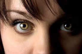
The functions of the eyes should not be overemphasized. We ought to thank God always for giving and keeping the members of our body complete. Below are some diseases of the eyes that we steemians should guide against.
💠STRABISMUS (Squint, Cross-eye)

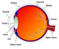
Strabismus is the inability of the eyes to move so that the same image fall on the corresponding part of the retina in both eyes. It is caused by exstraocular muscle weakness or defective nerve supply to the muscle, i.e., defective cranial nerve 111, IV or VI. In most cases the image falling on the squinting eye is suppressed by the brain, otherwise there is double vision (diplopia).
💠CATARACT
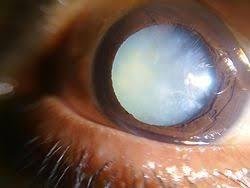
This is opacity of the lens which may be degeratative or congenital, bilateral or unilateral. In degenerative cataract there is gradual development of lens opacity that may be due to senile degeneration, X-rays, infrared rays (heat), trauma, diabetes, uveitis, and some drugs, e.g., long-term use of cortecosteroids, Senile cataracts usually developed in older aged groups. Other degenerative forms may developed at an earlier age depending on the primary cause. Developmental cataract may be due to genetic abnormality, e.g., Down's syndrome, or maternal infection in early pregnancy, e.g., rubella.
💠RETINAL DETACHMENT
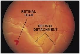
This occur when a tear or hole in the retina allows fluid to accommodate between the layers of retinal cells or between the retina and choroid. It is usually localized at first but as fluid collects the detachment spreads. There spot before the eyes, flashing lights due to abnormal stimulation of sensory cells, and progressive loss of vision. In many cases the cause is unknown but it may be associated with trauma yo the eyes or head ,tumours, haemophage, cataract surgery when the pressure in the eye is reduced or diabetic retinopathy.
💠RETROLENTAL FIBROPLASIA
This may occur in premature infants who receive oxygen therapy. The high oxygen tention in the blood may cause constriction of immature retinal arterioles. After oxygen therapy ceases they is disordered development of retinal blood vessels and the formation of fibrovascular tissue in the vitreous body causing varying degrees of interference with light transmission. Blindness may be cause by heamorrage in the vatreuos humour, detarchment of the or in very severe cases, by formation of opaque membranes behind the lens.
💠RETINITIS PIGMENTOSA
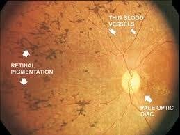
This us a hereditory disease in which there is degeneration of the retina, mainly affecting the rods. Defective vision in dim light usually becomes apparent in early childhood, leading to tunnel vision and eventually, blindness.
💠KERATOMALACIA
in this condition there is corneal ulceration, usually with secondary infection. This lacrimal glands and conjunctiva may be involved. It is cause by chronic vitamins A and protein deficiency in the diet. They may be softening or even perforation of the cornea. Night blindness (defective adaptation yo dim light )is usually an early sign of deficiency of vitamin A which is required for the reconstitution of rhodopsin(visual purple )after it has been exposed to light.
💠TUMOURS

(A). CHOROIDAL MALIGNANT MELANOMA
This is the most common ocular malignancy and it occurs mostly in older people. Vision is not usually affected until the tumour causes retinal attachment, usually when well advanced. The tumour spreads locally in the choroid, and blood-borne metastates develop mainly in the liver, bones, lungs and brain.
(B). RETINOBLASTOMA
This is a malignant tumour derived from embryonic retinal cells. It is usually evident before the age if 4 years and may be bilateral .it spreads locally yo the vitreous and nay grow along the optic nerve, invading the brain.
💠ACUTE DECRYOADENITIS
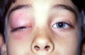
This is inflammation of the lacrimal gland ,usually unilateral .it may be due to spread of infection from the eyelids or surrounding structures, or be associated with measles ,mumps or influenza. The infection usually resolves but occasionally an abscess forms.
💠DACRYOCYSTITIS
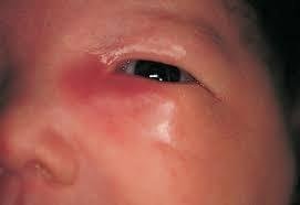
This inflammation if the lacrimal sac is usually associated with partial or complete obstruction of the lacrimal duct. In adults the blockage may be due yo narsal trauma, deviated nasal septum, nasal polyp or acute inflammatary nasal congestion.
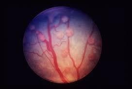
💠GLAUCOMA
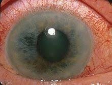
This is a group of conditions in which there Is increased intraocular pressure due to defective drainage if aqueous fluid through the canal of schlemm in the angle between the Iris and cornea in the anterior chamber .
🍥PRIMARY GLAUCOMAS
(A) CHRONIC OPEN-ANGLE GLAUCOMA
There us a gradual painless rise in the intraocular pressure with the progressive loss of vision. Peripheral vision is lost first but may not be noticed until only central (tunnel) vision remains. As the condition progresses, atrophy of the optic disc occurs leading to irreversible blindness. It is commonly bilateral and occurs mostly in people over 40 years of age. The cause is not known but there is a familial tendency.
(A) ACUTE CLOSED-ANGLE GLAUCOMA
This is most common in people over 60 years of age and usually affect one eye. During life the crystalline lens gradually increase in size, pushing the Iris forward .in dim light when the pupils dilates the lax iris bulges still further forward, and may come into contact with the cornea and close the canal of schlemm, raising the intra-ocular pressure. An acute attack may abort if the Iris responds to bright light, constricting the pupil and releasing the pressure on the canal. After repeated attacks spontaneous recovery may be incomplete, leading to further increase in intra-ocular pressure accompanied by severe pain, headache, nausea and vomiting. The cornea becomes oedematous and opaque and vjsionbis impaired.
🍥SECONDARY GLAUCOMA
The most common primary disorder is uveitis with the formation of adhesions between the Iris and lens capsule that obstruct the flow of aqueous fluid from the posterior to the anterior chamber. Other primary conditions include intraocular tumours, enlarged cataracts, central retinal veins occlusion ,intraocular haemorrhage, trauma to the eye.
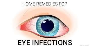
💠INFLAMMATION
(A) STYE (HORDEOLA)
This is pyogenic infection of sebaceous or meibomian glands of the eyelids margin. A 'crop' of styes may occur due to localized spread to adjacent galnds. Infection of meibomian (tarsal) glands may block their ducts, leading to cyst formation (chalazion) which may damage the cornea. The most common infecting organism is staphylococcus aureus.
(B). BLEPHARITIS
This is miceobila or allergely inflammation for the eyelid margins. The most common causes are stalhylococcal in fact I on or allergy to dandruff. If ulceration occurs, healing by fibrosus may distort the eyelid margins, preventing complete osure of the eye. This may lead to drying of the eye, conjunctivitis and possibly corneal ulceration.
(C) CONJUNTIVITIS
This is microbial or allergic inflammation. Microbial infection is usually by staphylococci, streptococci, pneumococci, herpes zoster (shingles), herpes simplex or measles viruses, Genacoccal infection may be acquired by the baby at birth if the mother is infected. Infection may spread to the cornea, causing ulcerayion and patches of opacity when the ulcers heal. Allergic conjunctivitis may be a comication of other autoimmune desease or be caused by a wide variety of antigens encountered in the environment, e.g., dust, pollen, fungus spores, animal dander, cosmetics, hair sprays, soaps.
(D) TRACHOMA
This is a chronic inflammatory condition caused by clamydia trachomatis in which fibrous tissues forms in the conjunctiva and cornea, leading eyelid deformity and possibly blindness. It occurs mostly where there is poor hygiene, especially in the middle East Africa and South East Asia. The microbes are usually spread by flies, communal use of contaminated washing water, crossed infection between mother and child.
(E) CORNEAL ULCER
This is local necrosis of corneal tissue, usually associated with corneal infection (keratitis) following trauma, or infection from the conjunctiva or eyelids. The most common infection microbes are staphylococci, streptococci, pneumococci and herpes simplex viruses. Acute pain, photophobia and lacrimation interfere with sight during the acute phase. Exstention ulceration and healing by fibrosis cause opacity of the cornea and irreversible sight loss.
(F) INFLAMMATION OF THE UVEAL TRACK (IRIS, CILIARY BODY, CHOROID)
(i) ANTERIOR UVEITIS
Iridocyclytis (inflammation of Iris and ciliary body) is the common and it may be acute or chronic. The infection may have spread from the outer eye but in most cases the cause is unknown. There is usually moderate to severe pain, redness, lacrimation and photophobia. In severe cases adhersion forms between the Iris and lens capsule, preventing the circulation of aqeuous fluid in the posterior and anterior chambers. This may cause the lens to bulge and occlude the canal of schlemm, raising intraocular pressure.
(ii) POSTERIOR UVEITIS
Choriorentinitis is the more common conditions. It may be caused by spread of infection from the front of the eye or be secondary to a wide variety of systemic conditions, including rheumatoid arthritis, Reiter's disease, ulcerative colitis, brucellosis. Complications that may occur include retinal detachment due to accomodation of inflammatory educate, secondary glaucoma, cataract.


Very educative
I'm grateful
👍
Boss man
Keep up the good work
Very educative. Good work sir
Highly educative and informative post. Keep it up bro
Alright
Thanks
Very good. Thank you
Cheers!!
Taking care of our eyes is good
It's really good to take care of the eyes. Thank you for your comment have followed you.
Eye is indeed the facial beauty of human
Are you an optician?
No sir
I'm a physiologist
Thank you for your comment
Incredible, keep it up...
Congratulations! This post has been upvoted from the communal account, @minnowsupport, by Fidelmboro from the Minnow Support Project. It's a witness project run by aggroed, ausbitbank, teamsteem, theprophet0, someguy123, neoxian, followbtcnews, and netuoso. The goal is to help Steemit grow by supporting Minnows. Please find us at the Peace, Abundance, and Liberty Network (PALnet) Discord Channel. It's a completely public and open space to all members of the Steemit community who voluntarily choose to be there.
If you would like to delegate to the Minnow Support Project you can do so by clicking on the following links: 50SP, 100SP, 250SP, 500SP, 1000SP, 5000SP.
Be sure to leave at least 50SP undelegated on your account.
Very informative and a little bit scary for me.