My Senior Psychology Thesis: Degree of (Intracerebral) Calcification and Calcification-Related Dysfunction
Degree of (Intracerebral) Calcification and Calcification-Related Dysfunction
Alex Van Aken
FGCU
29 November 2016
Introduction
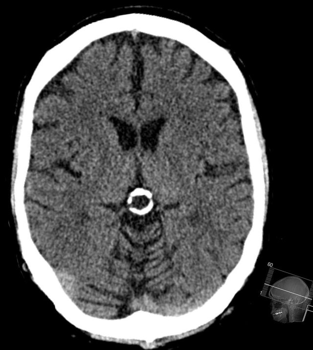
This paper is designed to review the current literature regarding brain calcifications, their sizes and locations, and how they may be playing a role which is rather large and over-looked in the medical world today. In many cases, when a doctor sees calcifications in the brain, it is typically calcification of the Pineal Gland (PG). In fact, one study found that 70% of their subject pools who have brain calcifications have Pineal Gland Calcifications (Yalcin et al 2016), making the pineal gland the most likely part of the brain to calcify, probably due to the region’s lack of a blood-brain barrier (Gaillard & Jones, 2010). There are definitely some things that need to change about modern calcification-assessment, because the main protocol that most doctors follow when they look at a CT scan, MRI or an X-Ray, is to look at the calcification of the PG (which is usually present as a white mass in the middle of the brain), and to determine whether this calcified mass is “pushed” off to either side. If it is displaced from its position in the center, the doctor can then be almost certain that there is a tumor on the enlarged side of the brain, which is displacing the PG, pushing it out of the way, over to the side (Fan 1983). Even though it has been determined that calcifications in the brain grow worse with advanced-age (Sigurdardottir et al, 2016) (Turgut et al 2008), there may be a lot more going on with regards to calcifications of certain areas of the brain. For example, there has been a statistically significant correlation found between Pineal Fluoride levels and Pineal Calcium levels. Fluoride accumulates in this remarkably small gland in the highest concentrations in the body (Luke, 2001).
When discussing this, however, it is vitally important to keep in mind that severity of the condition (physical size of calcified masses compared to non-calcified tissue) is the main defining variable being studied. This variable is typically referred to as the Degree Of Calcification (DOC). This is typically studied by Computed Tomography today (Turgut et al 2008) (Kitkhuandee et al 2014) (Yalcin et al 2016) or MRI (Lv et al 2010) (Sigurdardottir et al, 2016) however, before this technology was available, it was first assessed via X-Ray photograph by Schuller in the 1950’s (Yalcin et al 2016) and it is still detected via X-Ray photograph to this day (Bayliss et al 1985) (Mohammed et al, 2016). It is also noteworthy that CT scanning seems to be the most accurate way to assess prevalence and degree of calcification (Sedghizadeh, Nguyen, & Enciso, 2012).
It is also important to note that there are potentially multiple different types of calcification, that which is layered, and that which forms needle-like crystals (Humbert & Pevet, 1995). The locations, sizes and shapes of these calcifications are all important to look at.
The overall aim of this paper is to propose a paradigm of assessing this phenomenon of brain-calcification, the main variable concerned being the Degree Of Calcification (DOC) (size of calcified mass in millimeters), as well as any potentially negative consequences which could arise from having these calcifications, which will be referred to as Calcification-Related Dysfunction (CRD).
The main proposition to be made is that there is most likely a positive correlation between DOC and CRD, meaning, the more severely calcified the brain, the more possible calcification-related dysfunction could be occurring. The small niche of scientists who have been pioneering pineal gland and calcification-related intracranial phenomena are small in numbers, so due to the lack of current research, this subject needs to be researched further in order to determine whether DOC can be a potential causer of CRD. First, the broad topic will be discussed, then more specific potential connections will be addressed, including potential CRD as well as the various parts of the brain that can calcify over time.
Physiology
To understand the complexity of this situation, it is vital to understand the sizes of certain spaces and areas in the brain, in comparison to certain formations of calcification. The Pineal Gland is a small endocrine gland on the dorsal part of the mid-brain, and over time, this gland can actually calcify, looking like a small bone in the brain. This calcification can occur both on the outside of the PG as well as inside (involving the Parenchymal cells), and in the nearby-area of the pineal gland, which is actually quite small and tight. There is really no room inside this central area of the brain, especially for a large, hard, useless mass in the brain, which potentially displace other cells, cut off blood-flow, or even compress the cerebral aqueduct, which is right below the pineal-tectum area of the brain (Gaillard & Jones, 2010). It is vitally important that we find out what could potentially be wrong with this gland calcifying, because it is a major supplier of Melatonin and Serotonin for the body (Kunz et al 1998) (Kunz et al 1999).
Headaches & Migraines
Given that certain calcifications in the brain can build up over time, growing larger and larger, it is potentially a causation for many peoples’ headaches/migraines/head pressures/cranial congestion. Via CT Scan, calcifications in the brain can potentially displace brain tissue, potentially causing many of the uncomfortable “headache” criteria many people experience frequently. Potentially, when these calcifications grow larger, that mass pushing outwards, against brain tissue, could potentially produce a painful sensation in the head. There is a positive correlation between calcification of the PG and migraines (Ozlece et al 2015).
Hemorrhages
Certain calcifications in the brain, such as that of the PG, can become so large and obtrusive that they can actually potentially cause infarctions and hemorrhages in the brain by rubbing/scraping up against certain blood vessels in the brain. Calcifications have been correlated positively with intracerebral hemorrhaging (Kitkhuandee et al 2014).
Tumors
There is also a very high co-morbidity of both Pineal Gland Calcification (PGC) and Pineal Gland Tumors (Gaillard & Jones 2010), as well as PGC and cancers in the rest of the body (Drexler, 1963). There are also many types of tumors and cysts that can form on and around the general area of the pineal gland, potentially compressing the midbrain (Gaillard & Jones, 2010). In one study, an alarming majority of the tumors examined were originating from the PG (Lv et al 2010).
Insomnia
Since the mainstream science world has more or less accepted the Pineal Gland to be one of the major organs concerning the circadian rhythms, it is most certainly possible that Degree of Calcification (DOC) of the PG could play a role in the PG’s ability to produce sufficient Melatonin levels, resulting in healthy sleep and circadian rhythms. Number of healthy, active, uncalcified PG cells has actually been positively correlated with Melatonin by-product (aMT6s). In turn, this potential deficit of Melatonin could be leading to many of the sleep disorders we see today. In some cases, DOC increases with symptoms of Daytime tiredness, sleep disturbance, and stress (Kunz et al 1998) (Kunz et al 1999).
Bipolar Disorder & Depression
Since indole hormones like Serotonin and Melatonin are produced in large quantities in the pineal gland (Paltsev et al, 2016), it is possible that a decline in number of healthy pineal parenchymal cells could lead to a lack of endogenous serotonin, thus leaving a person prone to things like depression, bipolar disorder, or other conditions which could be a result of Calcification-Related Dysfunction (Gaillard & Jones, 2010).
Alzheimer's Disease
In a recent study, it has been shown that some alzheimer’s patients have the lowest number of active, uncalcified PG cells (pinealocytes) (Mahlberg, 2008). It is also noteworthy that Melatonin & Serotonin levels in the pineal begin to drop with old age (Paltsev et al, 2016). Alzheimer’s disease could potentially be accelerated due to DOC and any potential CRD.
Shizophrenia
There is also a very large occurrence of PGC amongst many Schizophrenics, and there could be an underlying connection between DOC, CRD and schizophrenia (Yalcin et al, 2016) (Sandyk, 1992).
Damaged Sense of Direction
Sense of direction is a very important adaptation that we have. Without it, we have a much lower level of self-efficacy. In a world where people have already gotten used to the convenience of having a GPS device at all times, it is important to keep in mind a sense of direction, as a survival instinct. PGC has been linked to a statistically significant decrease in sense of direction (Bayliss et al, 1985). This is a very important sense which has not yet become vestigial, however PG health most likely plays a role.
Widespread Calcification
It is interesting to note that the PG isn’t the only brain structure prone to calcification. It is, however, the most frequently-calcified part of the brain, and co-occurs with many other types of brain calcifications (Yalcin et al, 2016) (Daghighi et al, 2007). The most common intracranial calcification locations are: The Pineal Body, the Choroid Plexus, the Habenular (which is right above the PG and thus sometimes thrown into the larger category pineal/habenular), the dural surface, and even the basal ganglia.
Pineal Body
The pineal gland is typically the most-common part of the brain to calcify. 71.6% (Yalcin et al, 2016) and 71% (Daghighi et al, 2007) of people in these subject pools had Pineal Calcifications.
Choroid Plexus
70.2% of one study subject pool had calcifications of the choroid plexus, making it the second-most-prevalent brain calcification. 77.4% of those subjects found to have Choroid Plexus Calcifications also had Pineal Calcifications (Yalcin et al, 2016). In another study, 66.2% of subjects had Choroid Plexus Calcifications (Daghighi et al, 2007).
Habenular
Habenular Calcifications (HCs), were shown to be prevalent at 19.2% (Yalcin et al, 2016), and of those subjects who had HCs, 93.9% also had PC. In another study, this was reported at 20% (daghighi et al, 2007). This makes sense, because the Habenular is located immediately above the PG.
Dura Matter
12.5% had some form of calcification in this study (Yalcin et al, 2016) of the medial dural surface, appearing like a solid white line in CT scans, sometimes spanning a majority of the length of the skull, looking like an extra bone or plate in the brain. The most commonly co-existing calcifications seen alongside Dural Calcifications (DCs) were Choroid Plexus Calcifications, which co-occurred at a rate of 78.4%.
Basal Ganglia
1.3% had Basal Ganglia Calcifications (BGCs), and 82.6% of those people also had PCs in one study (Yalcin et al, 2016) Surprisingly, this type of intracerebral calcification was more common among women, in contrast with the others (Yalcin et al 2016). In another study, .8% had basal ganglia calcifications (Daghighi et al, 2007)
Discussion
Given the lack of past research on this subject, most scientists who are privy with it would agree that it is a widely misunderstood and overlooked situation. After all, when doctors see a ping-pong sized sphere of calcified tissue in the middle of somebody’s CT, MRI, X-Ray, etc., the main thing they ask themselves is whether or not this large, unnecessary, and potentially dangerous mass should be there or not. Due to the implications of some of these findings and speculations, this is most certainly a topic that deserves more attention in the mainstream science world. This topic should definitely be elaborated upon via further research, and gradually, physicians world-wide will eventually be able to more properly detect and think critically regarding brain calcifications.
Research in this field does have it’s setbacks and limitations. The pineal gland is so deep in the brain, it is hard to study in living people without advanced neuro-imaging, or in dead people without autopsying. Also, there are certain levels of “noise” that can occur during certain radiology procedures which can potentially disrupt the levels of quality delivered by the scans. These machines have only been around for a short time and the world is just now starting to use them to research on this subject. Worldwide, more and more people are looking to radiology to assess things inside the head, and these calcifications come up very commonly. Typically, doctors don’t really pay that much attention to them, unless they are actively looking for a tumor, or unless they are actively studying calcifications themselves. However, gradually, the mainstream science world is thinking about this subject and learning more with each passing year. This subject is vitally important because the brain regions discussed within this topic are highly important in maintaining proper mental health.
Conclusion
It is critically important that this particular topic eventually, at the very least, get the attention of the people who can do something for people: their doctors. If more and more doctors begin seeing this as more than just “business as usual”, then there will be a larger scope through which we can view our full-spectrum mental and physical health. There could potentially be a massive underlying cause of many different mental conditions, and that causer could potentially be Degree Of Calcification (DOC), which could potentially lead to Calcification-Related Dysfunction, such as, lack of blood flow due to large calcification pushing up against blood vessels, lack of Melatonin or Serotonin biosynthesis by the cells of the pineal gland, due to a low level of ‘active’ (uncalcified) PG tissue, or some other over-looked possibilities. In short, DOC could potentially cause or worsen CRD(s), all of which producing a type of “snowballing” effect, leading to mental disorders, decline in health, and other potential consequences.
Works Cited
Bayliss, C. R., Bishop, N. L., & Fowler, R. C. (1985). Pineal gland calcification and defective sense of direction. British Medical Journal, 291(6511), 1758. doi:http://dx.doi.org/10.1136/bmj.291.6511.1758
Daghighi, M., Rezaei, V., Zarrintan, S., Pourfathi, H. (2007). Intracranial physiological calcifications in adults on computed tomography in Tabriz, Iran. Folia Morphol, 66, 115-119.
Drexler, J. (1963). The calcified pineal body and mammary carcinoma. Cancer, 16(12), 1616-1617. doi:10.1002/1097-0142(196312)16:123.0.co;2-0
Fan, K. (1983). Pineal Calcification Among Black Patients. Journal of the National Medical Association, 75(8), 765–769.
Gaillard, F., & Jones, J. (2010). Masses of the pineal region: Clinical presentation and radiographic features. Postgraduate Medical Journal, 86(1020), 597. doi:http://dx.doi.org/10.1136/pgmj.2009.087460
Humbert, W. & Pévet, P. Cell and Tissue Research (1995) 279, 565-573. doi:10.1007/BF00318168
Kitkhuandee, A., Sawanyawisuth, K., Johns, J., Kanpittaya, J., Tuntapakul, S., & Johns, N. (2014). Pineal calcification is a novel risk factor for symptomatic intracerebral hemorrhage. Clinical Neurology and Neurosurgery, 121, 51-54. doi:http://dx.doi.org/10.1016/j.clineuro.2014.03.019
Kunz, D., Bes, F., Schlattmann, P., Herrmann, W. (1998) On pineal calcification and its relation to subjective sleep perception: a hypothesis-driven pilot study. Psychiatry Research, 82, 187-191.
Kunz, D. (1999) A New Concept for Melatonin Deficit on Pineal Calcification and Melatonin Excretion. Neuropsychopharmacology, 21, 765-772.
Luke, J. (2001). Fluoride deposition in the aged human pineal gland. Caries Research, 35, 125-8.
Lv, X.- F., Y.- W. Qiu, X.- L. Zhang, L.- J. Han, S.- J. Qiu, W. Xiong, G. Wen, Y.- Z. Zhang, and J. Zhang. (2010) Primary Intracranial Choriocarcinoma: MR Imaging Findings. American Journal of Neuroradiology 1994-1998.
Mahlberg, R., Walther, S., Kalus, P., Bohner, G., Haedel, S., Reischies, F.M., Kühl, K.P., Hellweg, R., Kunz, D. (2008) Pineal calcification in Alzheimer's disease: An in vivo study using computed tomography Neurobiology of Aging, 29, 203 - 209
Mohammed, K., Boakye, E., Ismail, H., Geneus, C., Tobo, B., Buchanan, P., & Zelicoff, A. (2016). Pineal gland calcification in kurdistan: A cross-sectional study of 480 roentgenograms. PLoS One, 11 doi:http://dx.doi.org/10.1371/journal.pone.0159239
Ozlece, H., Akyuz, O., Ilik, F., Huseyinoglu, N., Aydin, S., Can, S., & Serim, V. (2015). Is there a correlation between the pineal gland calcification and migraine? European Review for Medical and Pharmacological Sciences, 19, 3861-3864. Retrieved October 11, 2016, from MEDLINE.
Paltsev, M., Polyakova, V., Kvetnoy, I., Anderson, G., Kvetnaia, T., Linkova, N., Paltseva, E., Rubino, R., De Cosmo, S., De Cata, A., & Mazzooccoli, G. (2016). Morphofunctional and signaling molecules overlap of the pineal gland and thymus: Role and significance in aging. Oncotarget, 7. doi:10.18632/oncotarget.7863
Sandyk, R. (1992) Pineal and habenula calcification in schizophrenia. International Journal of Neuroscience. 67, 19-30.
Sedghizadeh, P., Nguyen, M., and Enciso, R. (2012) Intracranial Physiological Calcifications Evaluated with Cone Beam CT. Dentomaxillofacial Radiology (2012) 41, 675-78.
Sigurdardottir, L., Markt, S., Sigurdsson, S., Aspelund, T., Fall, K., Schernhammer, E., Rider, J., Launer, L., Harris, T., Stampfer, M., Gudnason, V., Czeisler, C., Lockley, S., Valdimarsdottir, U., & Mucci, L. (2016) Pineal Gland Volume Assessed by MRI and Its Correlation with 6-Sulfatoxymelatonin Levels among Older Men. Journal of Biological Rhythms 31, 461-469.
Turgut, A., Karakaş, H., Özsunar, Y., Altın, L., Çeken, K., Alıcıoğlu, B., Sönmez, I., Alparslan, A., Yürümez, B., Çelik, T., Kazak, E., Geyik, P., and Koşar, U. (2008) Age-related Changes in the Incidence of Pineal Gland Calcification in Turkey: A Prospective Multicenter CT Study. Pathophysiology 15, 41-48.
Yalcin, A., Ceylan, M., Bayraktutan, O., Sonkaya, A., & Yuce, I. (2016). Age and gender related prevalence of intracranial calcifications in CT imaging; data from 12,000 healthy subjects. Journal of Chemical Neuroanatomy, 78, 20-24. doi:10.1016/j.jchemneu.2016.07.008