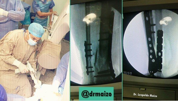
Osteosynthesis in orthopedic surgery
Osteosynthesis
Surgical joining of bone fragments by means of various (generally metallic) elements: wires, tapes, pins, plates and screws and intramedullary nails.
The material used must not cause any kind of irritation (chemical, mechanical or electrical) to the tissues, nor must it experience any phenomenon of osteolysis (neither primary nor secondary to electrolysis by use of metals of different composition).
The metals that meet these conditions are stainless steel, made up of iron and small quantities of carbon, chromium, nickel and molybdenum, and vitalium, a chromium alloy, cobalt and small quantities of nickel, molybdenum and tungsten. Titanium is currently widely used. It is also called internal fixation.

What's that all about?
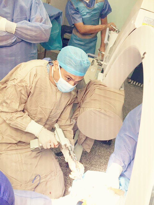
Internal fixation requires surgical exposure of the fracture site, open reduction of fragments and placement of a mechanical element to maintain reduction until complete healing. It has the advantage of producing a high degree of mechanical stability, its disadvantage being the associated surgical trauma.
The rigid conformation of the fixation limits the movement between the fragments to such a degree that there is no external callus formation, so it must be maintained for longer periods. It is necessary that the apparatus constitutes a whole with the damaged bone to resist the stress of physiological activity. They act based on the biomechanical principles of fragmentary compression, bridge and internal immobilisation.
Fragmentary compression holds the fragments together mechanically and can be static or dynamic. The first adheres the fragments, does not allow movement in the fracture focus with the physiological load and produces primary scarring, which in X-rays is seen as a gradual disappearance of the fracture line. Screws and cortical plates are good examples. In dynamic compression, the fixator transforms the physiological load into compression at the fracture site. This is how tension bands, containment or support plates, dynamic hip screws and unblocked intramedullary nails work.
An internal fixation element acts as a bridge when anchored in healthy bone proximal and distal to the fracture site, facilitating the transmission of physiological load from the proximal bone to the distal bone, without passing through the fracture site. As the fragments are not directly attached there is movement that translates into endostic and periosteal scarring. Intramedullary nails often act this way.

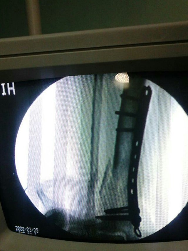
Plates:
There are different models, made of stainless steel or titanium, have several holes and are placed on the surface of the bones, secured with screws. Not all holes are necessarily used. They are classified on the basis of some of their attributes, either shape, design of the holes, site chosen for fixing or mode of application.
They are most frequently used in fractures of long bones, but also in arthrodesis of the spine and wrist. They require a larger surgical incision than other types of fixation. There is the possibility of alteration of cortical blood flow, due to the large contact surface, and consolidation, being able to reproduce the fracture when removing the plaque, by bone atrophy. In general, they base their operation on three biomechanical principles: dynamic compression, neutralization and containment or support. There are also some of special design.
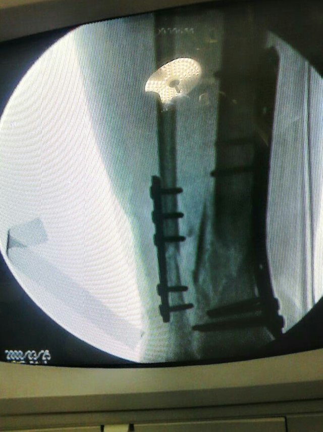
Compression plates compress the ends of the fracture, they are used to fix stable fractures maintaining reduction and compression. Compression can also be achieved through specially designed holes or by eccentric placement of the screws. They can be used in conjunction with fragmentary screws.
Dynamic compression plates (DCP), designed for axial compression, are one of the most commonly used types; they are recognized by their oval holes for the eccentric insertion of screws, whose walls are beveled towards the floor and inclined towards the medial. The weakest area of these plates is around the holes as this is the only area that can be bent.
The Low Contact or Impact Dynamic Compression Plate (LCP) is a newly developed type of compression plate, which differs from the PCD by the shape of the cut on its lower surface, which decreases the apposition surface around and between the screw holes, minimizing plate-periosteum compression, allowing greater capillary flow, and aiding the healing process. It has some degree of soft and elastic deformation, without concentrating stress around the holes.
Malleable reconstruction plates are widely used in pelvic, calcaneal and elbow fractures. They are pulled by the punching between the oval holes, which makes them easily malleable in the three planes, adapting to the shape and length required in complex bone surfaces.
Neutralization plates are placed on a comminuted fracture focus. Designed to protect the fracture surface, they transmit the forces of incurvation, torsion and axial load. They are often combined with fragmentary screws.
Containment or support plates are used in unstable fractures to support the thin periarticular cortical bone against compressive or axial loading forces, preventing its collapse. They are used in distal radius and tibial saucers.
Special design plates. The blade plate is used in supracondylic fractures. It has one of its ends with angles close to 900 to adapt to individual variations and with the shape of a chisel to be introduced into the metaphysis. The plate is fixed to the cortex with screws.
The specially designed anatomical plates are a wide range, designed for specific purposes: the 95° condylar plate for stabilization of fractures of the proximal and distal femur; the condylar support or containment plate for the distal femur; T 4.5 plate for the proximal humerus and tibia; 3.5 angled oblique T plate for the distal radius, etc.
If you need recommendations or help in orthopedic surgery and traumatology do not hesitate to contact me.
Dr. Leopoldo Maizo - Orthopedic Surgeon
If you want to read more I invite you to visit my page:
Firma diseñada por @themonkeyzuelans, contáctalos vía Discord "themonkeyzuelans#9087"
Great projects from the Steemit community:

- My Fundition campaign: https://fundition.io/#!/@drmaizo/6f88ggj8h


.png)
Always interesting. One thing I noticed from the picture is the c arm draping. Here it is required by law to cover the whole upper arm in plastic as well because it moves over the surgical field.
Takes good technique to drape that thing without contaminating one’s own gown. I always hated working right next to all that radiation !
Hi, you're absolutely right my friend @ceattlestretch, the C arm should be covered with great caution, something that not many radiology technicians agree with... Even the shields we use have been defeated for many years and do not adequately protect against radiation. Thank you for your great comment, my friend.
This project is being supported by @Fundition
Fundition is a next-generation, decentralized, peer-to-peer crowdfunding and collaboration platform, built on the Steem blockchain.
#upfundition and #fundition tags on Steem represent the projects that are started on https://fundition.io.
Are You Prepared to Make the World a Better Place too?
Read the full details of Fundition Fund program
Learn more about Fundition by reading our purplepaper
Join a community with heart based giving at its core