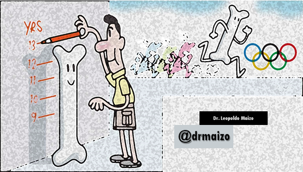
Bone development and formation
The beginning
The fetal skeletal system develops from mesenchymal cells and the neural ridge.
By the end of the third week of gestation, the paraxial mesoderm columns are segmented into mesodermal blocks called somites.
The cells of the neural crest migrate to the pharyngeal arches and form the bones and connective tissue of the craniofacial structures. Occipital somites and somtomers also help form the cranial vault and the base of the skull.
The somatic leaf of the lateral mesoderm intervenes in the formation of the bones of the extremities, the sternum and the pelvic and scapular waists.

Bones & Joints
Bones initially appear as condensations of mesenchymal cells that will constitute bone models. This condensation will delimit the beginning of the selective gene activity that precedes cell differentiation.
The flat bones develop in the mesenchyme inside pre-existing membranous sheaths, the mesenchymal models of the bones of the extremities are transformed into cartilage bone models, which will then carry out an endochondral ossification.
The cartilage appears during the fifth week. It arises from the condensation of mesenchyma, cartilage-forming cells, secrete collagen fibrils and the fundamental substance of the matrix. These fibers are then deposited in the matrix. Cartilage is divided into three types:
- Hyaline cartilage: wider distribution (e.g. in joints)
- Fibrocartilage (as in the intervertebral discs)
- Elastic cartilage (in the auditory pavilion)
Joints begin with the appearance of interzonal mesenchyma during the sixth week. Three types of joints are formed:
-fibrous: in which the interzonal mesenchyma is replaced by fibrous tissue (cranial sutures).
-Cartilaginous joints: where the interzonal mesenchyma is replaced by cartilage.
- synovial joints (such as in the knee).
The appendicular skeleton
The appendicular skeleton consists of pectoral waist, pelvic waist and bones of the extremities. They form during the fifth week as mesenchymal condensations that appear in the yolks of the extremities.
During the sixth week of development, the clavicle develops by ossification intramembranosa, at first, and then gives rise to growth cartilages at both ends. the pectoral waist and bones of the upper extremities appear before those of the pelvic girdle and lower extremities.
In the eighth week of development, primary ossification centers appear in the long bones and in the twelfth week they appear in almost all the bones of the extremities. The clavicles begin their ossification before the rest of the bones in the body, then the femurs.
Secondary ossification centres appear just before the birth of the bones, which form the knee joint. However, most secondary ossification centers appear after birth.
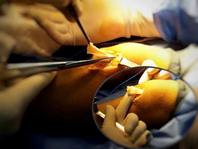
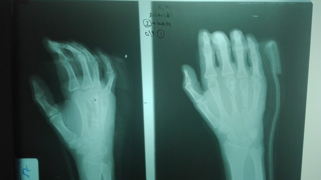
Dr. Leopoldo Maizo - Orthopedic Surgeon
Firma diseñada por @themonkeyzuelans, contáctalos vía Discord "themonkeyzuelans#9087"
Great projects from the Steemit community:

- My Fundition campaign: https://fundition.io/#!/@drmaizo/6f88ggj8h


.png)
This project is being supported by @Fundition the next-generation, decentralized, peer-to-peer crowdfunding and collaboration platform, built on the Steem blockchain.
Read the full details of Fundition Fund program
Learn more about Fundition by reading our purplepaper
Join a community with heart based giving at its core
Fundition is a non profit project, by supporting it with delegation you are supporting 200+ projects.
50SP100SP200SP500SP1000SP2000SP5000SP10000SP