ONCOLOGICAL CLINIC: SOFT PARTS SARCOMAS
Soft tissue sarcomas (SPB) are a large and heterogeneous group of rare tumors, which are characterized by frequently complex multidisciplinary treatment. Although in recent years there have been notable advances in the knowledge of the pathology and molecular biology of this disease, clinical treatment has evolved more discreetly and, in practice, mainly due to the rarity and complexity of this disease, the results are not always optimal.
• Alarm signs:
Tumors that affect soft tissue with any of the following signs should be considered potentially malignant:
Tumors greater than 5 cm.
Tumors that have experienced recent growth.
Deep (fixed) tumors.
Pain is not a symptom that per orients, but along with any of the above signs may reinforce the presumption of malignancy
• Diagnostic approach: Imaging studies, biopsy with radiological control.
Extremity sarcoma: Radiological explorations in the diagnosis and staging of soft tissue sarcomas of extremities. The method of choice in the diagnosis and local staging of an indeterminate or suspicious soft tissue mass of sarcoma is magnetic resonance imaging (MRI) with administration of intravenous contrast. In the event that MRI is contraindicated, a computed tomography (CT) with intravenous contrast should be used, preferably with additional reconstructions in sagittal and coronal planes. Valuations to include in the radiological report:
- Size
- Location (superficial or deep, compartmental or extracompartmental).
- Limits of the injury.
- Relationship with neurovascular structures.
- Extension of peritumoral edema.
- Contrast capture pattern.
- Suggestions of air to biopsy.
• Treatment of localized disease: Surgery of the primary tumor
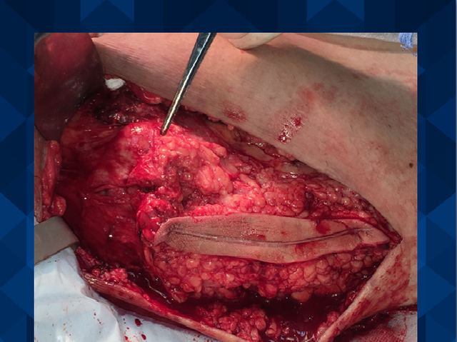
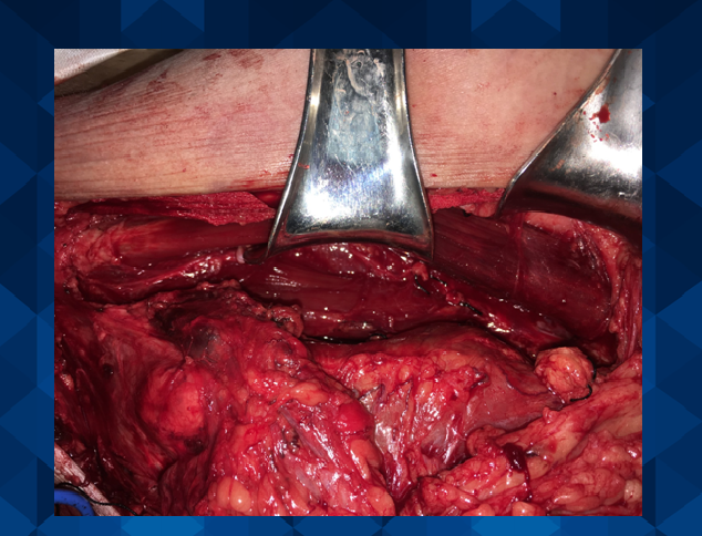
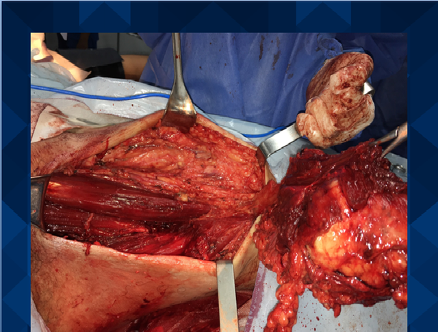
Surgery: Conservative surgery of the limb will be performed. Only the possibility of amputation will be considered when it is not possible to achieve wide margins and / or functional reconstruction of the limb. The main stages of conservative surgery are tumor resection, reconstruction and soft tissue coverage. The first phase is the oncological part of the procedure, which must be independent of the following phases and must never be influenced by them. The resection is performed according to the appropriate oncological surgical margins for each tumor. The margins are defined as: intralesional, marginal, broad and radical. The ideal margin must be broad or radical. A wide margin is considered to be one that is larger than 1 cm or that includes an undamaged anatomical barrier (muscle fascia, periosteum, perineurium). The reconstruction of the soft tissues, after the resection, is fundamental. If it is difficult or impossible, conservative surgery should be reconsidered and questioned. The implantation bed of the different types of reconstruction has to be adequate with good vascularization and adequate muscular coverage.
The techniques of plastic surgery, such as the transposition of the twin muscles, the dorsal muscle width, the free or pediculated grafts of skin or muscle-cutaneous, collaborate in the coverage of the different types of reconstruction and in the tension-free closure of the wound, which contributes significantly to the decrease in morbidity.
Here is a clinical case of soft tissue sarcoma that we received at an oncology surgery service:
PERSONAL INFORMATION
Name: Jairo Segundo Andrade Urdaneta
CI: 11,999,181
Age: 49 years old
Date and place of birth: 09/18/1967REASON FOR CONSULTATION
1.-Volume increase in the inner side of the left thighCURRENT ILLNESS
The patient is a 49-year-old male patient, natural and from the locality, with no relevant pathological history, who consulted for presenting an increase in volume on the inside of the left thigh for a month.
The patient reports having been operated by the Traumatology service in the month of July of the current year, sample is taken for BIOPSIABIOPSY
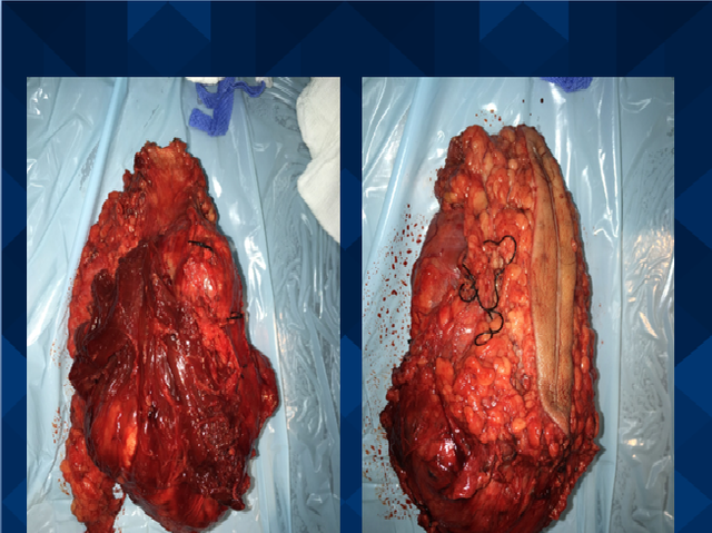
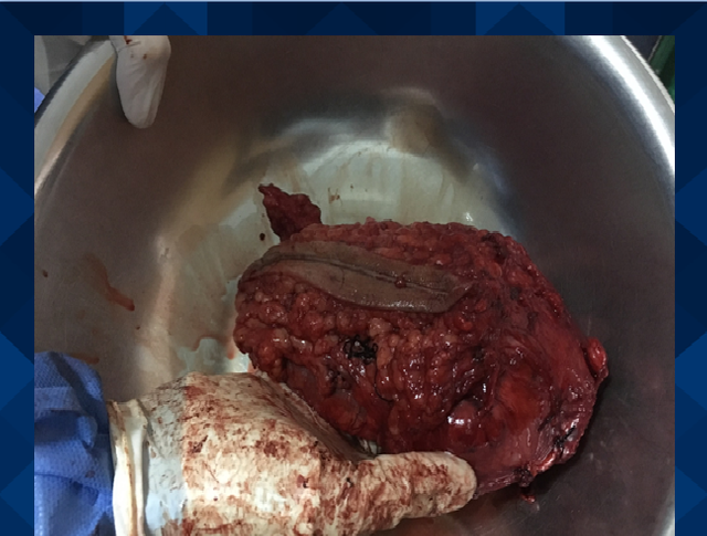
MACROSCOPIC REPORT:
Nodular tumor of 14x8x6cms is received, smooth external surface with hemorrhage zones, soft yellowish brown cut with areas of hemorrhage and other cystic mucinous contents. In the same container 2 fragments of muscle tissue of 3x2x1,5cms, soft light brown
MICROSCOPIC REPORT:
Histologically, malignant neoplasm of mesenchymal origin is observed, consisting of bundles of fusiform cells with large atypical hyperchromatic nuclei and mitosis, myxoid area and other necrosis. Normal muscle tissue
- DIAGNOSIS:
TUMORATION OF ADJUSTABLE COMPARTMENT OF THIGH LEFT:
- FUSOCELLULAR ARCHIVE WITH MIXOID CHANGES OF INTERMEDIATE DEGREE OF MALIGNITY
NOTE:
1.- Tumor of 14cms
2.- Grade II-III
Completely resected
Immunihistochemical studies are suggested to establish histogenesisIMMUNOHISTOCHEMISTRY
SAMPLE:
Left thigh tumor
IMMUNOHISTOCHEMICAL DESCRIPTION:
The Streptavidin-Biotin technique, the antigenic recovery method and adequate positive controls were used
ANTIBODIES PERFORMED IMMUNOEXPRESSION
Vimentina POSITIVE IN NEOPLASTIC CELLS
CD34 POSITIVE IN BLOOD VESSELS
Protein S100 NEGATIVE
Actina smooth muscle POSITIVE IN WALLS OF BLOOD VESSELS
IMMUNOHISTOCHEMICAL DIAGNOSIS:
Left thigh tumor
MALIGNANT FIBROHYSTIOCITOMA
- PHYSICAL EXAM
Weight: 77kg
Size: 1,80m
FC: 57 lpm
TA: 120/70 mmHg
FR: 16 rpm
Patient in regular clinical conditions, afebrile, hydrated, eupneic. Cardiopulmonary: audible vesicular murmur in both pulmonary fields without aggregates. Rhythmic heart sounds without murmurs. Abdomen soft, depressible not painful to superficial and deep palpation without visceromegalies. The tumor is palpated on the inside of the left thigh, approximately 4x3 cm in diameter. Neurological preserved.
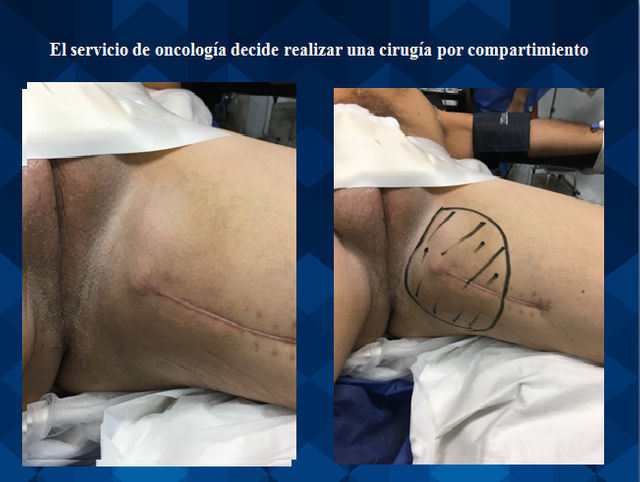
After continuous follow-up, and given the conditions, the oncology service decides to perform surgery by compartment

Thank God, satisfactory results were obtained and the patient was able to continue with his normal life.
I hope it will be of benefit and knowledge to you steemit community. Greetings, I hope to have your support :)
Congratulations @michelledayana! You have completed some achievement on Steemit and have been rewarded with new badge(s) :
Click on the badge to view your Board of Honor.
If you no longer want to receive notifications, reply to this comment with the word
STOP