Eyes of Scoliodon
They are also called photoreceptor organs.
Scoliodon has a pair of large eyes.
The eyes are located inside the eye sockets, one on each side of the head.
A. Eye muscle
The eyeball has an elliptical shape.
The eyeball remains attached to the inner wall of the orbit by 6 eye muscles and a cartilaginous stalk called the optic pedicle.
There are two groups of eye muscles. They are oblique muscles and rectus muscles.
There are 2 oblique muscles and they are the 1st oblique muscle lower and the 2nd oblique muscle upper.
The inferior oblique muscle is attached to the antero-ventral side of the eyeball.
The superior oblique muscle is attached to the anterodorsal side of the eyeball.
The action of the oblique muscles twists the eyeball.
There are 4 rectus muscles and they are 1. rectus inferior, 2. rectus superior, 3. rectus anterior and 4. rectus posterior.
The inferior rectus muscle is attached to the postero-ventral side of the eyeball.
The superior rectus muscle is attached to the postero-dorsal side of the eyeball.
The inferior and superior rectus muscles serve to rotate the eyeball in the vertical plane.
The anterior (or internal) and posterior (or external) rectus muscles are inserted anteriorly and posteriorly.
The anterior and posterior rectus muscles serve to rotate the eyeball in the horizontal plane.
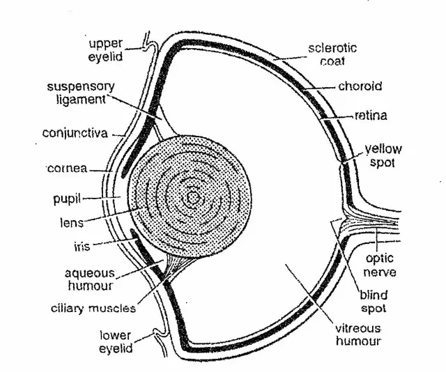
B. Internal structure
The outermost layer of the eyeball is the scleral layer.
The sclerotic layer is divided into two parts. The front part is the cornea and the back part is the sclera.
The cornea is transparent and allows light rays to enter the eye. It is covered by a thin and transparent conjunctiva.
The choroid layer is located below the sclerotic layer.
The choroid is a richly vascularized and pigmented layer.
At the anterior end of the choroid, just behind the cornea, the choroid forms a pigmented circular disc called the iris.
The iris has an opening in the center called the pupil. The entire eyeball is opaque to light except for the pupil.
Immediately behind the pupil is a large crystalline lens. The pupil cannot contract and dilate.
The suspensory ligaments are located just behind the iris and hold the lens in place.
The innermost layer of the eyeball is the retina.
This is the only part of the eyeball sensitive to light.
The retina consists mainly of elongated receptor cells, or rods, which are connected to the fibers of the optic nerve. Scoliodon has no cone cells.
A clear saline fluid, aqueous humor, fills a small chamber in front of the lens.
The very large chamber behind the lens is filled with a gelatinous substance, the vitreous. Scoliodon eyes.
C. Mechanism of vision/sight
The eyes of sharks are large but far apart, so binocular vision is not possible.
Power of accommodation is poor due to non-contracting pupils and little change in lens shape.
But the lens can be moved forward to capture more light.
Since the retina is devoid of cones, the fish is color blind and the eye cannot distinguish small details.
But the eye is adapted for close-up vision in the dark, and fish can see their prey.
Light rays pass through the cornea, pupil and lens and reach the retina.
The image formed by the retina is transmitted to the brain by means of the optic nerves.