Science Lesson: DNA (Part 6, How Does DNA Fit in the Cell?)
Science Lesson: DNA (Part 6, How Does DNA Fit in the Cell?)
Source
Preface
In previous posts we learned quite a bit about DNA, how information is stored, and how it is accessed by the cell to produce a desired product. We also learned about what DNA damage is, and some of the mechanisms by which the cell repairs these various damages. Those initial introductory blogs can all be accessed here:
Part 1-5: DNA Replication, Protein Expression, DNA Damage and Repair
Today we will discuss chromosomes, and how the enormous amount of DNA each our cells has, is compacted together to fit inside (our cells are pretty tiny after all)!
Break It Down For Me @justtryme90... How Much DNA DO We Have?
Well the human genome is around 3.3 x 10⁹ base pairs in length (That’s 3.3 Billion). The average length of a DNA base is around 3.4 Å long (Å = angstrom which is 1 X 10⁻¹⁰ m) This means that the human genome if stretched out end to end would be about 3.3 x 10⁻¹ meters in length, or about 33 centimeters! That’s actually quite long! Keep in mind that all of your cells have the very same amount of DNA inside of them. So with that in mind, also picture the size of a skin cell (the average skin cell is about 0.000030 m long or .0030 centimeters). How in the world do you fit 33 centimeters of DNA into a cell that is only 0.0030 centimeters long?! The answer to this lies into the structures that are formed to make up our chromosomes!
What is a Chromosome?
A chromosome is defined as a threadlike structure composed of both nucleic acids (DNA) and proteins found in the nucleus of most eukaryotic cells. Chromosomes are responsible for carrying the genetic information for the cell. You will notice in that definition that chromosomes are not just DNA! They also have a protein component and this is very important to how they are able to be so compacted!
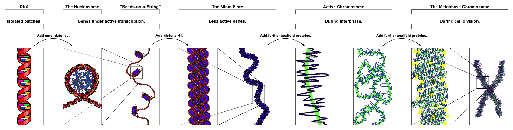
Figure 1: Overview of how a Chromosome is Assembled
As you can see from the above image you first have the DNA double helix, which is then wrapped around proteins called histone proteins into a ball shape called a nucleosome. These are then compacted together tighter and tighter until you end up with a thread like chromosome. Let us break these packing steps down a bit!
Histone Proteins and Nucleosomes
The first step in compacting DNA down into chromosomes involves interacting with a group of proteins called Histones. There are several of them (H2A, H2B, H3 and H4, the H stands for histone) and two copies of each one (So 8 total units) come together to form the histone complex (Figure 2 and 3):
Figure 2: Histone Complex (one side)
Source: Self with Use of Protein Database File 1KX5
Figure 3: Histone Complex (rotated 180°)
Source: Self with Use of Protein Database File 1KX5
The above figures are of an X-Ray crystal structure of the histone complex. One way scientists look at proteins is by getting them to form crystals under specific conditions, we then take the crystals and blast them with X-rays, which will diffract when they hit the crystal. This diffraction occurs in a pattern which can be observed, recorded and mathematically reassembled back into the original shape which caused the diffraction. Thus, we can actually see precisely what proteins look like (with extremely high resolution in some cases, 1.8 Å is the resolution of the above structure, and if you will recall that is smaller than the size of an individual nucleobase which makes it sufficient to see all bases and amino acids in the protein). In the above figures, I have colored each of the histone proteins in this complex (Red, Orange, Yellow, Green, Blue, Purple, Pink, Black) so you can see that there are indeed 8 separate proteins (2 copies of each of the 4 varieties of histone proteins). Around this complex DNA gets wrapped (approximately 147 base pairs worth of DNA), as you can see in Figure 4.
Figure 4: DNA Bound Histone Complex (Nucleosome)
Source: Self with Use of Protein Database File 1KX5
Compaction of Nucleosomes into 30 nm Fiber
After DNA is wrapped around the histones to form nucleosomes, they too get complexed together through the use of a 5th histone protein (H1). This complex is called a DNA fiber, and is about 30 nM in diameter (Figure 5).
Figure 5: The 30 nM Fiber
Finally Higher Level Packing
After formation of the 30 nm fiber (now technically a chromosome), there are a variety of not well understood scaffolding proteins that this fiber interacts with and allow it to continue to compact into the X shaped structure that you are familiar with as the chromosome (Figure 1). However you may find it interesting to know that this structure that you are so familiar with does not exist at all points in the cell cycle! In fact it only exists in a particular stage in a cells life during the process of cellular division called Mitosis.
Figure 6: Mitosis
Most of the time the chromosomes are just a jumbled mess of 30 nm fibers, however during prophase (Figure 6) is when the various scaffolding proteins begin to shape the fibrous DNA into even more compact shapes! These tightly compacted chromosomes continue to exist as the cell passes through metaphase, and separates into two daughter cells (anaphase). However once the mitotic process is complete, the DNA gets unpacked some again, to allow for protein expression and DNA replication to occur. The chromosome structure we all know and love is actually packed much too tightly for any of the DNA to be accessible for the cell!
Summary:
Human cells are full of an extremely massive amount of genetic material that through interactions with a group of proteins called histones, gets packed together into tight threads. These threads are what allow the DNA to effectively fit inside of the cell. During cellular division, chromosomes are further compacted through interactions with scaffolding proteins. These extremely compact structures are what most of us are familiar with (they have the X shape). However, they are packed together too tightly for the cell to utilize, so they only exist during this specific time in the cells life cycle.
Future Topics:
What is Cancer and what are its Causes?
Other Topics Suggested by You!
If you like my work, please consider giving me a follow:
Thank you for your support of my work!
Thanks :)
I hope you can also include, knowledge expanded a bit.
Awesome post ✅👍❤️🆙
Thanks! I appreciate the kindness!
Wishes you all the best.
that's where DNA methylation and histone acetylation come into play. looking forward to the next one!
Blockchain looks like DNA.
Excellent point! They are indeed very similar, I believe I made the analogy in one of my earlier posts in this very series!
The parallels between DNA and the block chain with regards to once something gets recorded its there to stay are strong.
Really cool post. One can follow and understand without being a specialist! :)
Thank you @lemouth, that is my goal! I want to make these complicated topics digestible for anyone who wants to learn!
Thank you for this brilliant series! #SteemitUniversity
Thank you for your kind words! I am happy you are enjoying it!
Great info thank you.
When I went to school (many years ago) we were taught that there were simple cells.
Now, if you give me the opportunity, I would like to teach you more about what is happening inside these cells.
This is why I always feel so wound up ;) as usual, great presentation. The repository is coming along nicely.
Thanks! I actually wrote this post because of one written by @renzoarg: https://steemit.com/science/@renzoarg/oedipus-this
In that article he discussed histone methylation a bit, and I thought it might be good to include in my background material just what histones were, and how DNA is involved with them. I could write a whole series on epigenetics actually. Its a really interesting, and relatively new field.
I remember recently reading about epigenetics and sexual orientation, it's a fascinating field.
I'm totally loving the reference you made to my article there :D
Your article is what inspired me to write this one. :)
Loved the article, I should check my feeds more often now that I'm following you.
A graphic addendum:
That video well encapsulates the things I have discussed throughout my posts! The DNA replication model they show applies to bacteria only however. It's known as the trombone model. Eukaryotic replication is a bit different. While the chromosomal packing material does not pertain to bacteria as they do not have Histones. However there are prokaryotic organisms (archaea) that do!