Cardiology #2 : The life-saving earphone, The Stethoscope Part 1
I bet many of the non-medical based personnel are curious about the most iconic symbol of physicians and doctors, the stethoscope. Trust me, before I join medschool, I have wondered numerous time about how this amazing instrument is capable of determining what kind of disease the person has. I have asthma and when I was younger, I had frequent visits to the emergency department because of its exacerbation. The doctors listened to my chest via the stethoscope and came out with treatment plans. Hence, I was intrigued about this magical device. What sort of sound does it produce? Does the stethoscope is computerized so it will tell the doctor the diagnosis directly? I used to think that the device works this way...
Stethoscope: "Bzz bzz, it's an asthmatic attack"
When I was in second year of med school, I have earned my very own stethoscope (yeay !) but clouds of disappointment came after that as I have no idea what am I listening to. Yes, it’s a freaking sound amplifier in which skills are needed to understand those noises. Oh yeah, try screaming at the disc-shaped structure and your ears will bleed profusely. Okay la, I’m not being literal.
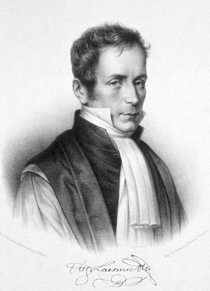
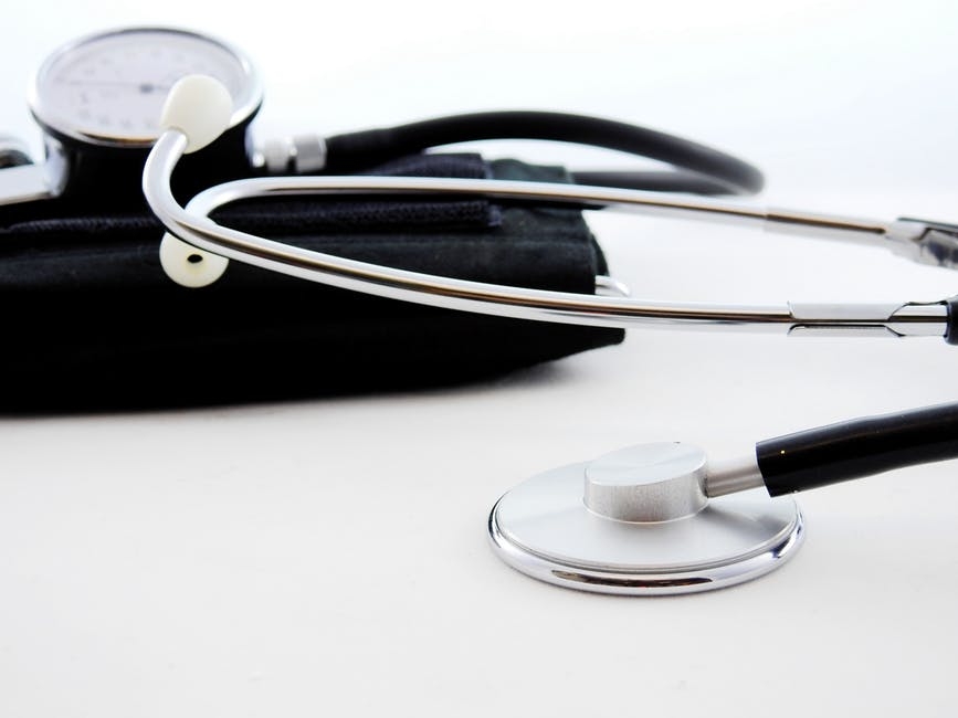
Stethoscope is a device that many doctors and nurses use to listen to sounds produced by the human body, except for loud noises projected through the mouth. Auscultation comes from the Latin verb Auscultare , which means “to listen”. A beautiful word huh, auscultare. Fret not, this device is just an amplifier. It doesn’t speak the diagnosis, but instead, experienced clinicians have the ability to interpret whatever that comes out from the earpiece. The earpiece is connected to the small-disc shaped resonator via two tubes. The act of listening to sounds generated from our body parts has originated way back among the Ancient Egyptions. A French physician with the name of René-Théophile-Hyacinthe Laennec , invented this miraculous device in 1816 in which he managed to diagnose numerous chest conditions. He devised this instrument when he finds that it is very uncomfortable to place his ears on a woman’s chest to hear heart sounds. He managed to conclude that there is possibilities to listen to chest sounds via a rolled piece of paper in between his ears and the patient’s chest, without requiring direct contact. I can’t imagine if doctors still place their ears on their patient’s chest nowadays…. Ironically, he succumbed to tuberculosis in 1826. Tuberculosis is a type of infection that prominently affect the lungs.
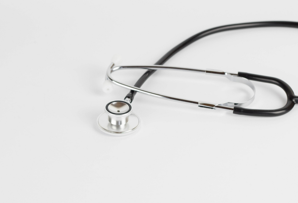
Even the most modern stethoscopes are simple, as they are consist of the diaphragm (larger, and flatter part of the chest piece), bell (smaller and more concave in shape) and the ear piece. You can switch this two parts of the chest piece (diaphragm and bell) by just twisting it. You’ll hear a click sound after a 180 degrees turn. The diaphragm excels in picking up high pitch sounds such as breath sounds and heartbeats. On the other hand, the bell is better in transmitting low pitch sounds, such as some heart murmurs (sounds of blood flowing) or bruits (a vascular murmur). The tubes is a Y-shaped configuration of rubber tubes that connects the earpiece to the diaphragm/bell. The tubing typically ranges from 45 to 68cm in length. The earpiece is made of small rubbers that cancels out external noise, similar to those structures in the earphones we use.
The surface area of the chest piece is much bigger than the tube. The chest piece vibrates when sound occurs. After that, these high frequency sounds travels to the ear piece in an amplified version.
Stethoscope helps to determine some abnormalities in breathing and heartbeat patterns, intestinal movements and blood flow. It can even aid us in detecting our blood pressure. Yeah, the digital one does not require any form of stethoscopes but doctors still use manual blood pressure measurement to reconfirm the blood pressure value especially when the result is too high or too low. Thus, the usual systems that are related include the cardiovascular, respiratory, and gastro-intestinal systems.
Cardiovascular System
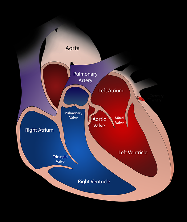
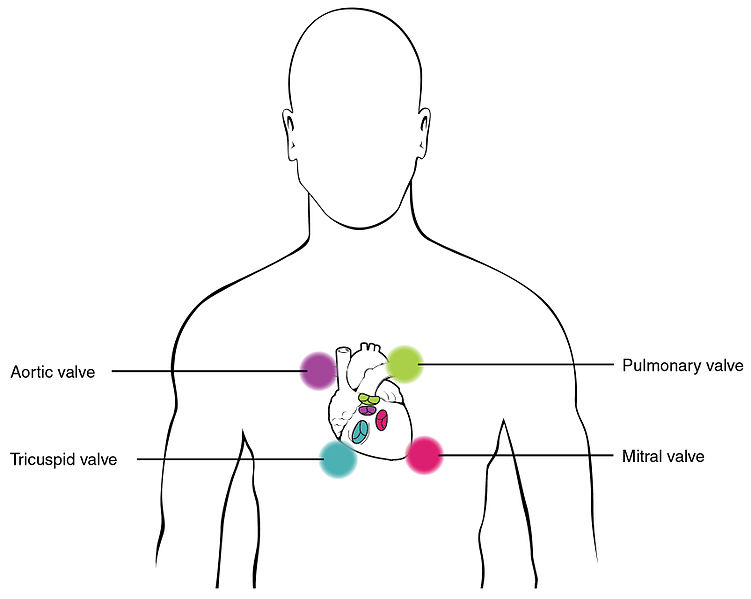
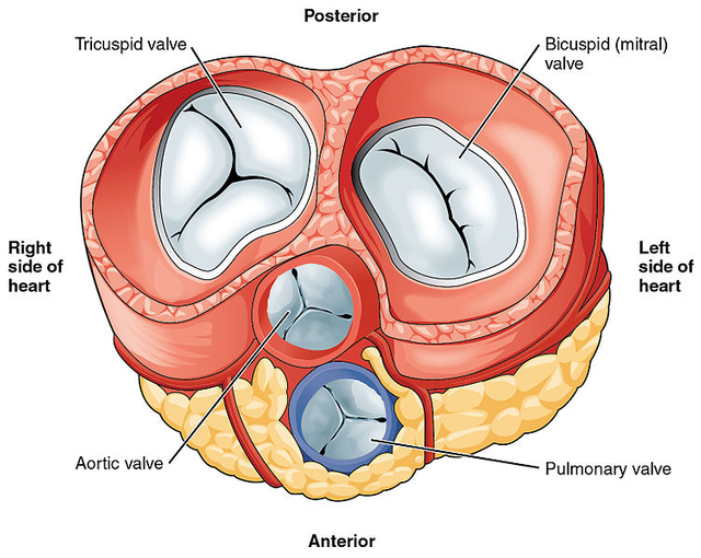
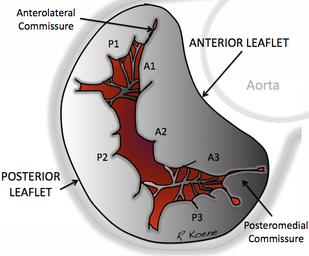
The stethoscope is able to pick up abnormal heart sounds of the heart. Here, both bell and diaphragm play important roles in amplifying those sounds so doctor can diagnose the condition that the person is having. There are four valves in the heart, the valves of the right side of the heart are the pulmonary and tricuspid valves and the left side consist of aortic and bicuspid (mitral) valves. Heart valves only allow single direction of blood flow and they are the separator of the atria from ventricles or the ventricles from the blood vessels. The valves are structurally made of cusps or leaflets. The mitral valve is the only valve with two cusps (hence, bicuspid) while the other valves contain three cusps. Heart murmurs are caused by turbulent blood flows especially when the heart valves are abnormal. The murmurs can be graded by the following scale:
• 1/6: Very faint, not always heard in all position.
• 2/6: Quiet, but not difficult to hear
• 3/6: Moderately loud
• 4/6: Loud + thrills (a vibratory sensation that can be felt on the skin)
• 5/6: Can be heard with the stethoscope partly off the chest + thrills
• 6/6: Can be heard with the stethoscope completely off the chest + thrills
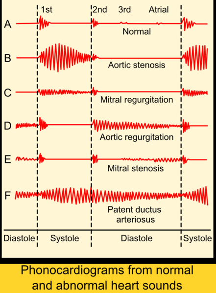
Systolic murmurs occur during the systolic phase (when the ventricles contract and ejects the blood out) and diastolic murmurs occur during the diastolic phase (when the ventricles are relaxed to allow blood to flow into it)
Mitral Stenosis
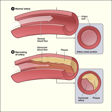
Stenosis means the abnormal narrowing of passage in the human body. Hence, this condition describes about the bicuspid valve being too narrow. This condition can result from previous rheumatic fever which is an infection of the heart caused by a B-hemolytic streptococcus. Inflammation of the valve area results in fusion and reduction in the mitral valve orifice area. The normal valve orifice (opening) is 4-6cm2 and severe stenosis is characterized when the orifice is less than 1cm2 . Symptoms include severe shortness of breath because the narrowing caused an increase in left atrial pressure and also the pulmonary (lung) circulation. Right heart failure also can occur in this process when the right heart needs extra effort to pump against the high pulmonary pressure. The symptoms are tiredness, abdominal distension and leg swellings. Due to the increased left atrial pressure, an opening “snap” will be heard to overcome the pressure. This is followed by a rumbling, mid diastolic murmur which is best heard via the bell when the patient is lying on the left side in expiration.
Mitral Regurgitation
Regurgitation in heart conditions happen when there is leakage of blood backwards. Mitral regurgitation causes left atrial dilatation. Similar to mitral stenosis, breathlessness and right heart failure symptoms can occur too. Mitral regurgitation can be present for many years before symptoms can happen as it is more insidious compared to mitral stenosis. Soft first heart sound can be heard due to the incomplete closure of the heart valves. Pansystolic murmur can be heard too. Pansystolic means throughout systole, thus the murmur heard is throughout the systolic phase. It is best heard via the diaphragm, loudest at the apex and radiates into the axilla (armpit area).
Aortic Stenosis
The aortic valve separates the right ventricle and the aorta. Aortic stenosis is the chronic progressive disease which commonly caused by calcifications of the leaflets. Individuals with this condition can present with exercise-induced syncope (loss of consciousness due to exercise). This happens because the blood cannot be ejected from the heart due to the severe narrowing of the aortic valve. Remember BP = TPR x CO? Check out one of my previous entry about Hypertension in which I have explained about it. Thus, the Cardiac Output (CO) is reduced which leads to low Blood Pressure (BP). Low BP causes the individual to loss his/her consciousness as there is lesser blood flow to the brain. The most obvious findings in Aortic Stenosis is an ejection systolic murmur that is usually diamond in shape (crescendo-decresendo). The murmur has a rough quality and best heard in the aortic area. The sound radiates to the carotid arteries.
Aortic Regurgitation
This condition can be caused by diseases affecting the aortic valve such as endocarditis (inflammation of the endocardium, a part of the heart) and diseases affecting the aortic root such as Marfan syndrome. Aortic Regurgitation is the leakage of blood from the aorta through the aortic valve, filling back the ventricles during diastole. Symptoms of Aortic Regurgitation includes breathlessness, chest pain, and pounding of the heart to the increased in left ventricular size in order to pump more blood out. Thanks to the leakage. On auscultation, there will be high-pitched early diastolic murmur best heard at the left sternal edge in the fourth intercostal space while the patient is leaning forward and breath held during expiration.
These are the common murmurs of the heart valves. There are others such as Tricuspid Stenosis/Regurgitation and Pulmonary Stenosis which are less common.
Bruit
Bruit, or vascular murmurs are abnormal sounds produced by turbulent flow in the arteries. This can be either due to a very high blood flow or when there is narrowing of the artery. Usually, doctors use Bell to listen to bruits. Most bruits occur during systole and it is pretty much dependent on the heart rate. So most arteries can generate bruits. Examples of arteries are Femoral, Renal, Carotid, and Aorta. Examinations of the cardiovascular system and the gastro-intestinal system are usually completed by searching for Carotid and Renal bruits respectively.
Alright, that’s all for Part 1 article of this blog. I will be discussing about the abnormal Breath Sounds as well as Bowel Sounds on my next article. Here's the summary of the murmurs pattern that can be heard upon auscultation. 1st indicates the first heart sound, and 2nd is the second heart sound. Lubdub lubdub lubdub.
References
- Kumar P., Clark M., Kumar & Clark's Clinical Medicine. Ninth Edition. London: ElSevier, 2017. Print
- What are the uses of stethoscope?. Healthfully. Retrieved on April 29, 2018, from https://healthfully.com/uses-stethoscopes-5104562.html
- The Stethoscope and How To Use It. Paul. Inside PA Training. Retrieved on April 29, 2018, from http://www.mypatraining.com/stethoscope-and-how-to-use-it/
- How Stethoscope Works. Julia Layton. howstuffworks. Retrieved on April 29, 2018, from https://science.howstuffworks.com/innovation/everyday-innovations/stethoscopes1.htm
- Stethoscope. Wikipedia. Retrieved on April 29, 2018, from https://en.wikipedia.org/wiki/Stethoscope
This is a masterpiece, am a 3rd yr optometry student and in my line of work we don't make us of the stethoscope more often as the other health professional but notwithstanding knowledge is precious and power so i really appreciate the indept delineations given. Keep it up bro, i will follow you so i get to read your subsequent posts.
Gosh thank you for the compliments. I’ve followed you too. Again, thank you 🙏🏽
I never knew it could be used for diagnosis like you explained. While growing up, I thought it was just to check if one is alive, don't mind me... I was fascinated by its use anyway
Hahaha yeap. This article is meant to those who are curious about the stethoscope. Thanks for reading !
You welcome
You've been upvoted by TeamMalaysia Community :-
To support the growth of TeamMalaysia Follow our upvotes by using steemauto.com and follow trail of @myach
Vote TeamMalaysia witness bitrocker2020 using this link vote for witness