Forearm fractures in children. Orthopedic conservative management.
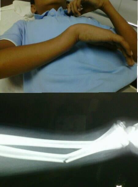
Image source: personal.
The bone of children is more flexible than the adult due to the water content in relation to the mineral component, also, the periostium (layer that covers the surface of the bone) is thicker and confers additional resistance to the shearing forces.
On the other hand, their ligaments are stronger, so that a trauma that can generate a sprain in the articulation of a mature skeleton, would cause a fracture in pediatric patients as it will happen in segments such as the ankle or wrist.
The power of recovery (osteogenesis) and remodeling is greater because the blood supply has characteristics that differ in the adult (it is metaphysial and extends to the physis) that is, it covers the body and the ends of the bone.
The most affected segments are the diaphysis (the middle of the long bone) and the physis (growth nuclei).
The main mechanism of injury is the fall with the hand in extension, however it can occur with the wrist in flexion, and depending on it the deformity of the forearm will be evidenced according to the forces generated by the muscles that originate or insert there. It is important to highlight the fact that many times these recurrent fractures may be due to metabolic or genetic diseases or child maltreatment.
The most affected age group is between 12 and 16 years, representing 45% of all fractures in this stage, with a higher prevalence in the male gender.
There are several fracture patterns depending on the mechanism of injury: warped or "green stalk", run, incomplete, complete. Etc.
In the case of plastic phenomena the bone suffers a bending without but the cortical are intacts) or incomplete fractures (when only one of the cortical injured) the treatment is conservative, obviously, it is imperative to perform an adequate anamnesis, physical examination, radiographs with the protocol projections: anteroposterior and lateral and once the diagnosis is confirmed, it is immobilized with a plaster cast, to relieve pain and edema for a period of approximately 4 weeks.
In the case of displaced fractures, the treatment consists of a closed reduction, ideally under sedation or general anesthesia according to the age of the patient, with subsequent immobilization by means of a closed brachiopalmar plaster, that is, covering all the upper limb, with the elbow in flexion of 90 ° can be in pronation, supination or neutral position depending on the level of the lesion with respect to the diaphysis. The neurovascular elements of the area (capillary refill, coloration, sensitivity) should always be monitored, considering that this reduction should be as anatomical as possible since the biomechanics of the forearm is very particular and does not admit inconsistencies or angulations greater than 20 °.
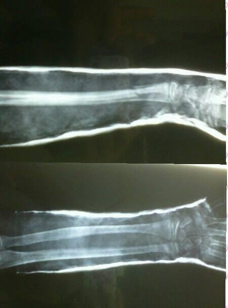
Image source: personal.
The patient and his / her parents must be explained that he / she must remain for 72 hours with the upper limb raised above the level of the heart, even when sleeping, to favor venous return, and avoid stasis, and therefore, a severe compression due to edema that merits plaster removal.
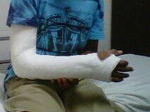
Image source: personal.
Regular radiological examinations should be performed to evaluate the status of the bone and rule out possible displacements, if so, another closed reduction is made and sometimes its treatment is surgical.
The plaster should be modified at 3 weeks, antebrachialpalmar, releasing the elbow joint to initiate mobilization , and according to the evolution of the formation of the bone callus, when the consolidation of the fracture is confirmed (between 4 to 6 weeks ) the cast is removed and rehabilitation therapy is initiated.
I want to conclude this article emphasizing that in Orthopaedics there are absolute and relative indications for surgeries, and more so in children, who have a capacity for amazing regeneration and remodeling.
Bibliographic references:
-Fracturas y luxaciones, Koval and Zuckermann, Ed. Marbán, 3era edición.
-Rockwood and Green´s. Fracturas en el niño. 5 ta edición. Madrid: Marbán, 2003
This post has been voted on by the steemstem curation team and voting trail.
There is more to SteemSTEM than just writing posts, check here for some more tips on being a community member. You can also join our discord here to get to know the rest of the community!
Thanks!!
Hi @soanna!
Your post was upvoted by utopian.io in cooperation with steemstem - supporting knowledge, innovation and technological advancement on the Steem Blockchain.
Contribute to Open Source with utopian.io
Learn how to contribute on our website and join the new open source economy.
Want to chat? Join the Utopian Community on Discord https://discord.gg/h52nFrV
Thanks!