How can the bioluminescence GFP from jellyfish be used in medical applications?
Many of us, if not all of us, must have seen images of brightly glowing cells in an array of colours being used as covers of scientific journals and biomedical test books; or the images of white lab mice glowing green under UV light, before. But have you ever paused for a moment to wonder the significance of this to medical research and applications? In a related case, you may have seen the term GFP being commonly used in scientific papers and books and maybe harbouring questions with regards to not only its meaning but also why and how scientists are able to make cells emit such arrays of beautiful luminescence even when GFP is not found in some of these cells naturally. Whatever is your situation, today's post will address all of that. It will be focusing on the meaning of GFP, its discovery and how scientists are manipulating it for applications in medical research.
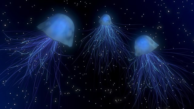
[Source: Pixabay. CC0 licensed]
What is GFP?
GFP is an acronym for green fluorescent protein. It is a florescent protein first isolated from the luminous organ of the jellyfish Aequorea victoria by a Japanese marine biologist Dr Osamu Shimomura in the early 1960s. When blue light is beamed on the creature, the molecules of GFP proteins absorb it, change its wavelength and emit a green light in response. This seemingly simple feature has continued to be an important tool in the study of cell biology following the isolation of its cDNA in 1992. The ease with which GFP can be introduced into cells and its characteristic ability to generate the intrinsic chromophore in the absence of cofactors or enzymatic components (i.e it does not need additional chemicals, enzymes or other substances to glow), have made it an indispensable tool for medication research.
The secret behind the usefulness of the GFP in medicine
Understanding the biochemical processes underlying the formation of proteins and the way they interact within biological organisms, is essential in fashioning appropriate, effective response in the event of malfunctioning of a vital component, yet these proteins are so tiny to be observed even under an electron microscope. No thanks to their clear or colourless nature which make observation of important structures inside a cell, an impossible task. Hence, being able to catch even a glimpse of these biochemical processes in action is so invaluable to medical researchers. The challenge in the observation and understanding of the important biochemical processes is, however, fast becoming a thing of the past because researchers have realized that they can attach GFP to virtually anything inside a cell, beam a blue light on the cell and then observe the activities and the movements of the GFP-bearing molecules.
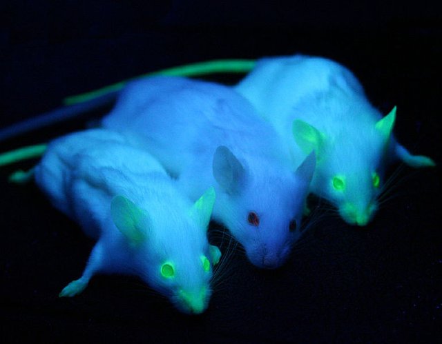
[GFP mice. Source: wikimedia commons. Author: Ingrid Moen et al. CC BY 2.0 licensed]
Already, this fact has been demonstrated in countless of scenarios since it was first demonstrated. Researchers have attached GFP to insulin-producing cells of the pancreas in studies which have helped them to understand how these cells are made with possibility of informing new diabetes treatments. GFP has equally been tagged to the sperm cells of fruit fly in a study that seeks to shed light on how certain sex cells maximize their chances of being fertilized. There were many reports in the 90s whereby geneticists introduced the jellyfish's GFP gene into mice as well as other laboratory animals which resulted in these animals having bodies luminous with GFP. This is so because the protein was expressed all over the animals' bodies including their muscle cells, brain cells and other organs. Now, scientists could watch how cancer cells spread in these animals or how their immune systems fight off viruses and other harmful intruders by shinning lights of appropriate wavelengths on them.
GFP allow scientists and researchers to visualize their experiments. It reveals what, perhaps, might have been impossible to visualize with naked or aided eyes in the form of glowing, bright colour. More so, it enables scientists to carry out experiments and observe them in live cells . Living Cells are examined under a microscope in real time and not as dead or attenuated cells.
With GFP, researchers can visualize where within a cell a protein is located. All they need to do is to attach GFP to gene of interest and then determine where this gene is coded when it is eventually expressed by the recipient organism. Again, researchers can label proteins with a visible marker and be able to observe the behaviors of such proteins under varying physiological conditions like a change in the expression of other genes or the introduction of a molecule. It is also possible to observe specific structures within a cell or specific cells within an organism through the application of light-emitting proteins. A good example of this is found in the field of connectomics using the "brainbow" mouse. Here, neurons within the mouse brain are marked with fluorescent proteins like GFP, allowing researchers to closely observe how these neurons interact with one another and thus providing insight into how mammalian nervous systems function.
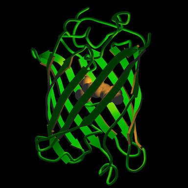
[Structure of GFP. Source: flickr commons. Author: NIH. CC BY-NC 2.0 licensed]
Following realization of the enormous applications of GFP and innovations in medical science, many color variants of the fluorescent protein have been isolated from many other organisms, such as fireflies and corals, and circulated throughout the world to provide colorful decoration to living cells during medical research. Today, they are not only being used as markers for protein localization in living cells, they are also being used to observe intracellular environments under varying physiological conditions. Suffice to say the applications of fluorescent proteins in medicine are simply inexhaustible; you can go here and here for more details on medical applications of GFP.
In conclusion, as has been noted above; the applications of GFP is so numerous to be fully captured in a single piece of blog article. For this reason, it is best stated that most (if not all) of the current biomedical research would be far less informative without them. Little wonder the 2008 Nobel Prize in Chemistry was awarded to the three scientists namely Drs Osamu Shimomura, Martin Chalfie, and Roger Tsien whose works led to the discovery and development of GFP; thus confirming the significance of this discovery and also the recognition accorded to its impact in biomedical research. Thanks for reading.
References
- Green florescent protein
- Deep sea creatures shed light on the future of medical imaging
- How a jellyfish protein transformed science
- From jellyfish to cancer cells: Why florescent proteins are crucial to biomedical research
Truly yours,
@sciencetech
STEM contributor.
Hello there @sciencetech :)
You really choose interesting topics to write and educate your readers about. For me, the piece of information about the use of GFP to obtain understanding into diabetes treatment is new and fascinating! I never realized how extensive the use of this protein is! Truly incredible.
You take care and thank you once again for sharing well-written, interesting content with us :)
Nature is beautiful :)
Indeed!
Thanks for finding the post interesting and particularly for being here. I do really appreciate this comment.
Thanks for finding the post interesting and particularly for being here. I do really appreciate this comment.
This post has been voted on by the steemstem curation team and voting trail.
There is more to SteemSTEM than just writing posts, check here for some more tips on being a community member. You can also join our discord here to get to know the rest of the community!
Hi @sciencetech!
Your post was upvoted by utopian.io in cooperation with steemstem - supporting knowledge, innovation and technological advancement on the Steem Blockchain.
Contribute to Open Source with utopian.io
Learn how to contribute on our website and join the new open source economy.
Want to chat? Join the Utopian Community on Discord https://discord.gg/h52nFrV
I started to do research into GFP and other bioluminescence for my masters project. In addition to GFP, I went down the rabbit hole of diatoms. Following the work of Pani Jurek with Living lamps, I met the biologist Stanisław Łoboziak, and from there I was hooked! He helped me to see the complex world of bioluminescense and to realize that our ancestors didn't really live in the dark, as we thought!
I should backtrack and say that the project for my masters was based in Matera, one of the oldest, continuously inhabited cities in the world. We have always thought that troglodyte culture--cave dwellers--lived in darkness, but it is probably more correct to say that they had bioluminescence to light their underground homes and produce oxygen!
Hmm.... maybe I should write about that? I never thought it would be of interest to anyone else. :)