Mitochondrial morphology, microscopy and - facepalm
Some six months ago, @sco organized a challenge. Steemit scientists got a call to submit the worst method they used with the appropriate tag #myshittiestmethod. The biggest trauma for the majority of participants was related to the countless protocols that included purification and extraction of various cell structures. All those protocols are dating back to the 60’s, the golden age of classical biochemistry.
The logic was relatively simple and straightforward: take a piece of plant/animal --> extract cellular structures (usually 2 days of hard work) --> treat that extract as yet another “pure chemical” --> measure give to a slave student to do a lot of kinetics.
If you feel burning desire to do some of those methods, I’ll give you the references:
• Johnson, Diane, and Henry Lardy. "[15] Isolation of liver or kidney mitochondria." Methods in enzymology. Vol. 10. Academic Press, 1967. 94-96.
• Lyons, James M., and John K. Raison. "Oxidative activity of mitochondria isolated from plant tissues sensitive and resistant to chilling injury." Plant Physiology 45.4 (1970): 386-389.
• Sussman, M. R. "Molecular analysis of proteins in the plant plasma membrane." Annual review of plant biology 45.1 (1994): 211-234.
The next age was the age of probes. Now the experimental pathway was: take something alive-ish --> feed it with a probe (don’t kill it) --> and put it in a spectrometer or under a microscope
• Bačić, Goran, and Miloš Mojović. "EPR spin trapping of oxygen radicals in plants: a methodological overview." Annals of the New York Academy of Sciences 1048.1 (2005): 230-243.
• Dugan, L. L., et al. "Mitochondrial production of reactive oxygen species in cortical neurons following exposure to N-methyl-D-aspartate." Journal of Neuroscience 15.10 (1995): 6377-6388.
Back to the XXI Century, let’s do some math and molecular biology
A few days ago, my colleague showed me the paper that describes subtle changes of the mitochondrial structures that reflects stress. Paper was published in 2017, and there is a high chance you missed it as it has relatively modest 15 citations.
Key ingredients for this experiment were:
• Cell culture
• MitoTrackerRed CMXRos, excitation in the range between 520 and 600 nm, emission in the range between 580 and 680 nm
• mEmerald-Mito 7 plasmid, excitation: 487 nm, emission: 509 nm
• Microscope from the shad, with some appropriate filters, Ex/Em (450-490 / 500-550 nm) and (540-552 / 590-660 nm)
Now it’s the time to do some math!
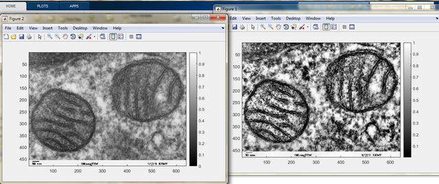
Fortunately, there is a relatively user-friendly ImageJ plug-in Mitochondrial Network Analysis (MiNA) also available on GitHub https://github.com/ScienceToolkit/MiNA.
The key elements of the algorithm include the enhancement of local contrast (CLAHE). Contrast-Limited Adaptive Histogram Equalization (CLAHE) is also available as Matlab function or as macro for Fiji.
CLAHE is related to the technique of histogram equalization. “One image is worth 1000 words”, so let me explain you by showing the example and it will be clarified in a second.
The image is from Wiki, the rest was done by me.
As you can see, the Gaussian distribution of intensities became “a rectangle”.
Adaptive histogram equalization, AHE, computes local histograms instead of only one, global histogram. As you can assume, it will be great for the local enhancement especially on the boundaries between objects and background. Unfortunately, it will be terrible in uniform regions, because the noise will be amplified.
The most advanced method CLAHE, uses the local distribution around the pixel to estimate if the region is uniform or not and choosing the appropriate transformation function.
I’ve tried to find some significant modifications, but there is nothing significantly improved after the first introduction of CLAHE.
There is yet another approach I’ve found for mitochondria, and it was described in this article and here, also followed by software and https://github.com/vianamp/MitoGraph .
Described method is very suitable for examination of confocal images (Z-stacks).
If you want to find more approaches and appropriate fluorescence probes, check this Review.
The results and aftermath
Now there is no doubts that the internal structure of mitochondria can be changed due to the response to oxidative stress, in Alzheimer dissease, or the presence of *OH.
If the boss asks you to do some old-fashioned isolation, on Monday speak loudly what you read in this post and get fired by Thursday!
Instead of telling you Goodbye: “Mitochondria is a powerplant of cell!”


I like the golden age of biochemistry. But I only need to read about it, not do it :D
Lucky you and lucky me because I've drifted from it long time ago. Poor students...
This post has been voted on by the steemstem curation team and voting trail.
There is more to SteemSTEM than just writing posts, check here for some more tips on being a community member. You can also join our discord here to get to know the rest of the community!
Hi @alexs1320!
Your post was upvoted by utopian.io in cooperation with steemstem - supporting knowledge, innovation and technological advancement on the Steem Blockchain.
Contribute to Open Source with utopian.io
Learn how to contribute on our website and join the new open source economy.
Want to chat? Join the Utopian Community on Discord https://discord.gg/h52nFrV