How Painkillers Like Aspirin Look Under the Microscope
“Crystals that will ease your pain!”
Almost everyone is familiar with painkillers, and most of us have taken them. But have you ever wondered about how they would look underneath a microscope? Of course your question at the time taking that painkiller would have been “will it ease my pain?”
There are a large number of painkillers available from the weakest aspirin to the strongest oxymorphone. Each works in a different way. Most people only need to take painkillers for a few days or weeks at most, but some people need to take them for a long time.
Painkillers can be taken by: mouth as liquids, tablets, or capsules, by injection, or via the rectum for example, suppositories. And some are even available as a creams or an ointment.
So why take micrographic pictures of pain medication?
Behind every used painkiller there is a story. Stories of the people taking their pain medication, but most of these stories are of course no happy stories. One day a few years back, I did not have a happy story, and I was bound for a long time taking strong pain medication.
During this period I was not really able to do my normal photography work. So I found back some old moleskins, and went trough all my notes. One think popped-up several times “Micrographs”. Combining my “two” passions; science and photography.
As an licensed medical laboratory analyst, I saw lots of beautiful things underneath the microscope when I was working at the RIVM. Capturing these moments in a form of art was always a wish.
So I started to build a setup that would enable me to go back to this “happy place”. Getting to know the world in a different way, by using things we “consume” in our “daily” life, but putting them underneath a microscope. Creating images “from another world” with a different perspective. Where structures, shapes, patterns, details, colours an many other things will (hopefully) make you look astonished.
After a lot of research about possibilities (within the available budget), I found my starting setup. A Novex-B microscope that was able to show me some of the worlds within microscopy.
By “modding” this microscope I could use the techniques like; bright-field, cross polarised, dark-field, phase contrast and oblique illumination.
A few weeks later the setup arrived, and the first thing that popped-up in my mind, was to try and see if I can make the medicine I was taking visible. But unfortunately that first step of making the particular medication visible by trying to crystallise it failed.
So where to start?
Like the introduction almost everyone has taken some painkillers in there life, so we can all relate to these medicine. The most common OTC (over the counter) pain medications are aspirin, acetaminophen, ibuprofen, diclofenac & naproxen. So starting with these 5 painkillers would be a good start.
The First results In the last months I have been experimenting allot to get the best results in therms of how to crystallise and capture these 5 OTC pain medications. I can happily report that I have managed to get beautiful micrographs of aspirin, acetaminophen & diclofenac.
Diclofenac:
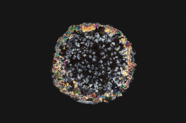
Diclofenac crystals after waiting for 72 hours, made visible by using a cross polarised light microscope.
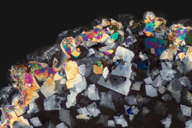
Diclofenac crystals after waiting for 72 hours, made visible by using a cross polarised light microscope. (100% zoom of above image)
Acetaminophen:
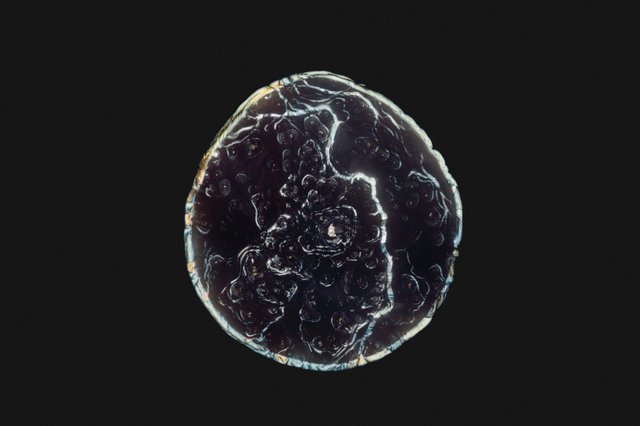
Acetaminophen crystals after waiting for 3 hours, made visible by using a cross polarised light microscope.
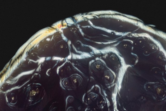
Acetaminophen crystals after waiting for 3 hours, made visible by using a cross polarised light microscope. (100% zoom of above image)
Aspirin:
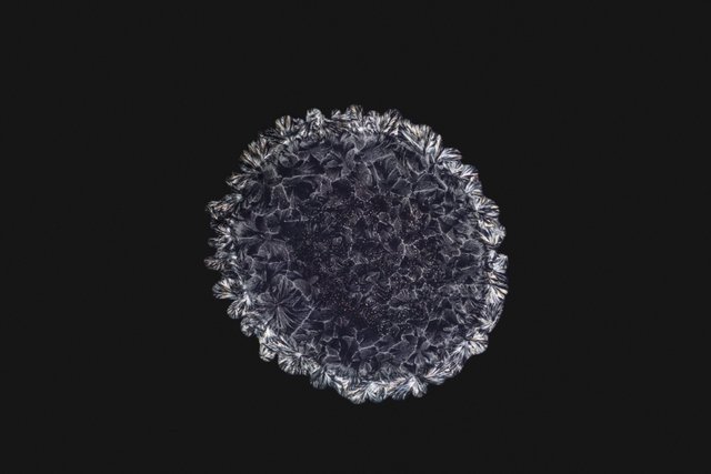
Aspirin crystals after waiting for 1 hour, made visible by using a cross polarised light microscope.
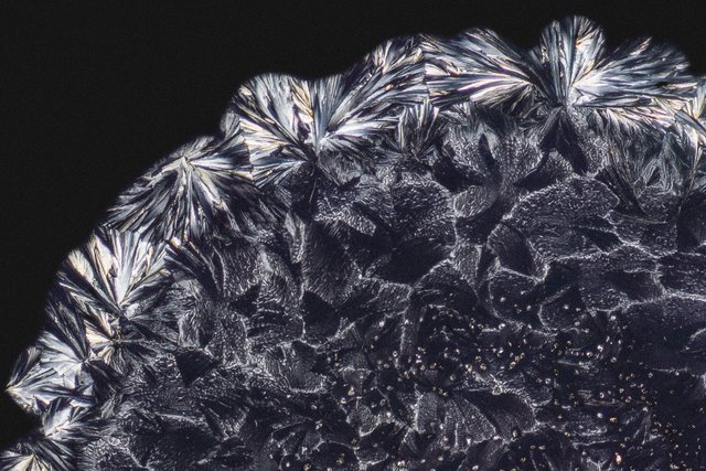
Aspirin crystals after waiting for 1 hour, made visible by using a cross polarised light microscope. (100% zoom of above image)
So next time you take one of the world’s most popular painkillers aspirin, diclofenac or acetaminophen, envision this microscopic molecule working its magic in your body.
#micrograph #photography #art #science #microscope #education #technology
Wow, I love seeing creative posts like this. You have a ton of potential "material" for future posts. You also have a unique advantage, as not too many people have access to the equipment needed to capture these beautiful images. It sort of reminds me of microscopic photos of grains of sand, flowers, sea shells, etc. It always amazes me how things photographed at that level look similar to very large things i.e. images captured from space. Great job. Looking forward to more pictures. What items can we look forward to in the future?
Thank you Cotough for your response. Yes, the world is I think still so unexplored, and new microscopic techniques are developed and used since the first microscope from 1632.
Lucky now for the general public this also means that they can now buy there own microscope for a fraction of the price it costed years ago. Even mobile phones are nowadays turned into small cheap microscopes, or what about the x$ "cardboard microscope". Amazing, since this also made it more feasible for me to start creating micrographs from home.
Even tho we have seen al lot trough microscope and there are a crazy amount of scientifically papers on them. Unfortunately there are also a lot of things that are not really visualised yet or are hard to find on high resolution. This is something I would like to cover from the perspective of the things we encounter and consume in our daily live combining it with the strength of the stories that come with it.
Some of the items you can look forward to in the future are:
1.) Turning medicine intake in to stories; meaning that there are millions of people in the world that are treated by medicine. And all these unique people have there own story. By visualising there crystalised medicine under a microscope I hope to tell there unique stories. For example someone that is taking insuline, cant live without. Imagine that I would be able with this project to visualise there medicine with right equipment (still need to invest allot of time and money to get everthing I need to do a brought spectrum of medicine crystlisations). This would give the person more interaction with its medicine (maybe this helps the person mentally). Next to that he or she is able to share its story with the world. This by a visual representation of its medicine creating more awareness for his or here situation. Since form experience people are really attracted to these images. Maybe the use of insuline or being a diabetic is already accepted, but there are a lot of deceases and medications that are much more difficult to talk about (taboo) (for example AIDS). Imagine giving those people a chance to tell there story more openly about how they got it when they find a positive connection with the viewer of the art form.
2.) Visualising individual stories by imaging tears underneath a microscope, showing and capturing unique and genuine stories of the donators.
3.) turning images in to hight mappings and turn them in to 3D printed objects that can be placed in open spaces for people to interact with.
4.) Explaining more about processes, techniques of the microscope and its capturing proces.
5.) Seeking interaction with a new audience on Steemit, and looking for collaborations between different kinds of intrest.
There are so many things I would like to do and write about, the only problem at the moment is to find the time and budget to get a lot of these ideas to work.
It kinds of makes me think and reflect about how atoms change behavior when we observe them....and of coarse HOW we observe them.